Explore
 Validate
 Learn
Learn Western blot
Antibody data
- Antibody Data
- Antigen structure
- References [5]
- Comments [0]
- Validations
- Western blot [1]
- Immunocytochemistry [3]
- Other assay [3]
Submit
Validation data
Reference
Comment
Report error
- Product number
- MA5-13817 - Provider product page

- Provider
- Invitrogen Antibodies
- Product name
- IGF1R alpha Monoclonal Antibody (24-60)
- Antibody type
- Monoclonal
- Antigen
- Other
- Description
- MA5-13817 targets IGF-1 Receptor alpha in IA, ICC/IF, WB and IP applications and shows reactivity with Human and Mouse samples.
- Antibody clone number
- 24-60
- Concentration
- 0.2 mg/mL
Submitted references Diane-35 and Metformin Induce Autophagy and Apoptosis in Polycystic Ovary Syndrome Women with Early-Stage Endometrial Carcinoma.
Analysis of Increased EGFR and IGF-1R Signaling and Its Correlation with Socio-Epidemiological Features and Biological Profile in Breast Cancer Patients: A Study in Northern Brazil.
IGF-1R nuclear import and recruitment to chromatin involves both alpha and beta subunits.
Low levels of insulin-like growth factor type 1 receptor expression at cancer cell membrane predict liver metastasis in Dukes' C human colorectal cancers.
Aurintricarboxylic acid induces a distinct activation of the IGF-I receptor signaling within MDA-231 cells.
Liu Y, Wang Y, Yao D, Chen X, Zhang F, Feng Y, Li X
Genes 2022 Jan 12;13(1)
Genes 2022 Jan 12;13(1)
Analysis of Increased EGFR and IGF-1R Signaling and Its Correlation with Socio-Epidemiological Features and Biological Profile in Breast Cancer Patients: A Study in Northern Brazil.
Silva Rocha F, da Silva Maués JH, Brito Lins Pereira CM, Moreira-Nunes CA, Rodriguez Burbano RM
Breast cancer (Dove Medical Press) 2021;13:325-339
Breast cancer (Dove Medical Press) 2021;13:325-339
IGF-1R nuclear import and recruitment to chromatin involves both alpha and beta subunits.
Mills JV, Osher E, Rieunier G, Mills IG, Macaulay VM
Discover. Oncology 2021;12(1):13
Discover. Oncology 2021;12(1):13
Low levels of insulin-like growth factor type 1 receptor expression at cancer cell membrane predict liver metastasis in Dukes' C human colorectal cancers.
Nakamura M, Miyamoto S, Maeda H, Zhang SC, Sangai T, Ishii G, Hasebe T, Endoh Y, Saito N, Asaka M, Ochiai A
Clinical cancer research : an official journal of the American Association for Cancer Research 2004 Dec 15;10(24):8434-41
Clinical cancer research : an official journal of the American Association for Cancer Research 2004 Dec 15;10(24):8434-41
Aurintricarboxylic acid induces a distinct activation of the IGF-I receptor signaling within MDA-231 cells.
Haimsohn M, Beery R, Karasik A, Kanety H, Geier A
Endocrinology 2002 Mar;143(3):837-45
Endocrinology 2002 Mar;143(3):837-45
No comments: Submit comment
Supportive validation
- Submitted by
- Invitrogen Antibodies (provider)
- Main image

- Experimental details
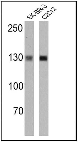
- Western blot analysis of IGF-1 Receptor alpha was performed by loading 25 µg of SK-BR-3 (lane 1) and C2C12 (lane 2) cell lysates onto an SDS polyacrylamide gel. Proteins were transferred to a PVDF membrane and blocked at 4ºC overnight. The membrane was probed with an IGF-1 Receptor alpha monoclonal antibody (Product # MA5-13817) at a dilution of 1:100 overnight at 4°C, washed in TBST, and probed with an HRP-conjugated secondary antibody for 1 hr at room temperature in the dark. Chemiluminescent detection was performed using Pierce ECL Plus Western Blotting Substrate (Product # 32132). Results show a band at ~155 kDa.
Supportive validation
- Submitted by
- Invitrogen Antibodies (provider)
- Main image
- Experimental details
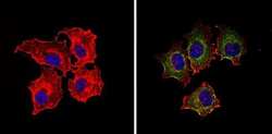
- Immunofluorescent analysis of IGF-1 Receptor alpha (green) showing staining in the cytoplasm and membrane of MCF-7 cells (right) compared to a negative control without primary antibody (left). Formalin-fixed cells were permeabilized with 0.1% Triton X-100 in TBS for 5-10 minutes and blocked with 3% BSA-PBS for 30 minutes at room temperature. Cells were probed with an IGF-1 Receptor alpha monoclonal antibody (Product # MA5-13817) in 3% BSA-PBS at a dilution of 1:20 and incubated overnight at 4 ºC in a humidified chamber. Cells were washed with PBST and incubated with a DyLight-conjugated secondary antibody in PBS at room temperature in the dark. F-actin (red) was stained with a fluorescent red phalloidin and nuclei (blue) were stained with Hoechst or DAPI. Images were taken at a magnification of 60x.
- Submitted by
- Invitrogen Antibodies (provider)
- Main image
- Experimental details
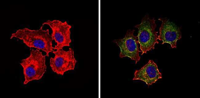
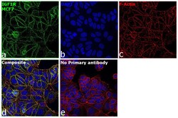
- Immunofluorescence analysis of Insulin-like growth factor 1 receptor was performed using 70% confluent log phase MCF7 cells. The cells were fixed with 4% paraformaldehyde for 10 minutes, permeabilized with 0.1% Triton™ X-100 for 15 minutes, and blocked with 2% BSA for 1 hour at room temperature. The cells were labeled with IGF1R alpha Monoclonal Antibody (24-60) (Product # MA5-13817) at 1:100 in 0.1% BSA, incubated at 4 degree celsius overnight and then labeled with Goat anti-Mouse IgG (H+L) Highly Cross-Adsorbed Secondary Antibody, Alexa Fluor Plus 488 (Product # A32723), (1:2000), for 45 minutes at room temperature (Panel a: Green). Nuclei (Panel b:Blue) were stained with ProLong™ Diamond Antifade Mountant with DAPI (Product # P36962). F-actin (Panel c: Red) was stained with Rhodamine Phalloidin (Product # R415, 1:300). Panel d represents the merged image showing Membrane localization. Panel e represents control cells with no primary antibody to assess background. The images were captured at 60X magnification.

- Submitted by
- Invitrogen Antibodies (provider)
- Main image
- Experimental details
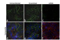
- Knockdown of Insulin-like growth factor 1 receptor was achieved by transfecting MCF7 cells with Insulin-like growth factor 1 receptor specific siRNA (Silencer® select Product # S7213, S7211). Immunofluorescence analysis was performed on untransfected MCF7 cells (panel a,d), transfected with non-specific scrambled siRNA (panels b,e) and transfected with Insulin-like growth factor 1 receptor specific siRNA (panel c,f). Cells were fixed, permeabilized, and labelled with IGF1R alpha Monoclonal Antibody (24-60) (Product # MA5-13817, 1:100) followed by Goat anti-Mouse IgG (H+L) Highly Cross-Adsorbed Secondary Antibody, Alexa Fluor Plus 488 (Product # A32723), (1:4000). Nuclei (blue) were stained using SlowFade® Gold Antifade Mountant with DAPI (Product # S36938), and Rhodamine Phalloidin (Product # R415, 1:300) was used for cytoskeletal F-actin (Red) staining. No signal was observed in siRNA slide of specific signal was observed upon siRNA mediated knockdown (panel c,f) confirming specificity of the antibody to Insulin-like growth factor 1 receptor (Green). The Images were captured at 60X magnification.

Supportive validation
- Submitted by
- Invitrogen Antibodies (provider)
- Main image
- Experimental details
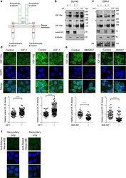
- Fig. 1 Full length IGF-1Ralpha- and beta-subunits are detectable in the nucleus of prostate cancer cells a Domain structure of mature IGF-1R at the plasma membrane, showing extracellular alpha-subunit (green), transmembrane beta-subunit (red), extracellular (ED), transmembrane (TM), kinase domains (KD) and disulphide bonds, modified from []. b , c Western blots showing detection of both IGF-1Ralpha- and beta-subunits in the nucleus of DU145 ( b ) and 22Rv1 ( c ) cells. Cells were serum-starved for 24 h, treated with solvent (4.5 uM HCl) or IGF-1 (50 nM) for 20 min before fractionation. All western blots shown are representative of 3 independent biological repeats. Arrowheads: 220 kDa IGF-1R proreceptor. d Images showing immunofluorescence staining of IGF-1Ralpha and IGF-1Rbeta in DU145 cells. DU145 cells were serum-starved and IGF-treated as in b-c followed by immunofluorescent staining. Graphs: mean +- SD nuclear IGF-1Ralpha- and beta-subunits (n >= 30 cells per condition for n = 3 biological repeats). Data were analysed by unpaired student''s t test and quantification of nuclear levels of IGF-1Ralpha- and beta-subunits showed a significant increase on treatment with IGF-1 (*p < 0.05; **p < 0.005; ***p < 0.001; ****p < 0.0001) e Images showing IGF-1Ralpha and IGF-1Rbeta immunofluorescence in DU145 cells. Cells were grown in full medium with 10% FCS and incubated with solvent (0.1% DMSO) or IGF-1R inhibitor BMS-754807 (100 nM) for 6 h at 37 degC and processed for immunofluore

- Submitted by
- Invitrogen Antibodies (provider)
- Main image

- Experimental details
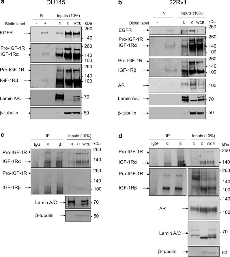
- Fig. 2 Nuclear IGF-1Ralpha- and beta-subunits originate from the cell surface and interact in the nucleus a, b Western blots showing detection of cell surface biotinylated IGF-1R in the nucleus of DU145 ( a ) and 22Rv1 ( b ) cells. Detection of cell surface biotinylated EGFR in nuclear fractions acts as a positive control. Western blots shown are representative of 3 independent biological repeats. c, d Co-immunoprecipitation Western blot showing interaction of IGF-1Ralpha- and beta-subunits in the nucleus of DU145 ( c ) and 22Rv1 ( d ) cells. Western blots shown are representative of 3 independent biological repeats
- Submitted by
- Invitrogen Antibodies (provider)
- Main image
- Experimental details
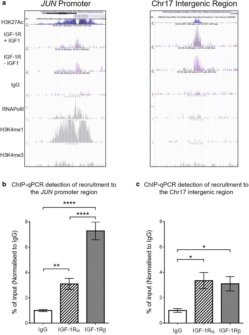
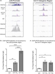
- Fig. 3 Recruitment of IGF-1Ralpha is detectable at IGF-1Rbeta DNA binding sites. a Images from the UCSC genome browser of previously obtained ChIP-seq data showing IGF-1R binding peaks that denote recruitment of RNAPolII and IGF-1Rbeta to the JUN promoter and IntChr17 [ref ]. b , c Quantification of recruitment of IGF-1Ralpha or IGF-1Rbeta vs IgG negative control to IGF-1Rbeta binding regions, detected by ChIP-qPCR. Results represent mean +- SEM of triplicate independent experiments, each with 3 technical replicates. Recruitment of both IGF-1Ralpha and IGF-1Rbeta vs IgG was detected at the JUN promoter ( b ) and putative IntChr17 enhancer (c; p* < 0.05, p** < 0.005, p**** < 0.0001, Tukey''s multiple comparisons test)