Explore
 Validate
 Learn
Learn Western blot
 Immunocytochemistry
Antibody data
- Antibody Data
- Antigen structure
- References [9]
- Comments [0]
- Validations
- Immunocytochemistry [1]
- Immunohistochemistry [2]
- Blocking/Neutralizing [1]
Submit
Validation data
Reference
Comment
Report error
- Product number
- AF-305-NA - Provider product page

- Provider
- R&D Systems
- Product name
- Human/Mouse IGF-I R/IGF1R Antibody
- Antibody type
- Polyclonal
- Description
- Antigen Affinity-purified. Detects human and mouse IGF-I R/IGF1R in direct ELISAs and Western blots. In direct ELISAs and Western blots, 25-50% cross-reactivity with recombinant mouse IGF-I R is observed. In direct ELISAs, less than 1% cross-reactivity with recombinant human IGF-II R is observed.
- Reactivity
- Human, Mouse
- Host
- Goat
- Conjugate
- Unconjugated
- Antigen sequence
P08069- Isotype
- IgG
- Vial size
- 100 ug
- Concentration
- LYOPH
- Storage
- Use a manual defrost freezer and avoid repeated freeze-thaw cycles. 12 months from date of receipt, -20 to -70 °C as supplied. 1 month, 2 to 8 °C under sterile conditions after reconstitution. 6 months, -20 to -70 °C under sterile conditions after reconstitution.
Submitted references Determination of Mammalian Target of Rapamycin Hyperactivation as Prognostic Factor in Well-Differentiated Neuroendocrine Tumors.
Antibody-Drug Conjugates Bearing Pyrrolobenzodiazepine or Tubulysin Payloads Are Immunomodulatory and Synergize with Multiple Immunotherapies.
Mycobacterium leprae-induced Insulin-like Growth Factor I attenuates antimicrobial mechanisms, promoting bacterial survival in macrophages.
Transplantation of Human Neural Progenitor Cells Expressing IGF-1 Enhances Retinal Ganglion Cell Survival.
LMP1 promotes expression of insulin-like growth factor 1 (IGF1) to selectively activate IGF1 receptor and drive cell proliferation.
IGF-IR mediated mammary tumorigenesis is enhanced during pubertal development.
FGFR2 is amplified in the NCI-H716 colorectal cancer cell line and is required for growth and survival.
Hyperactivation of the insulin-like growth factor receptor I signaling pathway is an essential event for cisplatin resistance of ovarian cancer cells.
Differential expression of receptor tyrosine kinases (RTKs) and IGF-I pathway activation in human uterine leiomyomas.
Lamberti G, Ceccarelli C, Brighi N, Maggio I, Santini D, Mosconi C, Ricci C, Biasco G, Campana D
Gastroenterology research and practice 2017;2017:7872519
Gastroenterology research and practice 2017;2017:7872519
Antibody-Drug Conjugates Bearing Pyrrolobenzodiazepine or Tubulysin Payloads Are Immunomodulatory and Synergize with Multiple Immunotherapies.
Rios-Doria J, Harper J, Rothstein R, Wetzel L, Chesebrough J, Marrero A, Chen C, Strout P, Mulgrew K, McGlinchey K, Fleming R, Bezabeh B, Meekin J, Stewart D, Kennedy M, Martin P, Buchanan A, Dimasi N, Michelotti E, Hollingsworth R
Cancer research 2017 May 15;77(10):2686-2698
Cancer research 2017 May 15;77(10):2686-2698
Mycobacterium leprae-induced Insulin-like Growth Factor I attenuates antimicrobial mechanisms, promoting bacterial survival in macrophages.
Batista-Silva LR, Rodrigues LS, Vivarini Ade C, Costa Fda M, Mattos KA, Costa MR, Rosa PS, Toledo-Pinto TG, Dias AA, Moura DF, Sarno EN, Lopes UG, Pessolani MC
Scientific reports 2016 Jun 10;6:27632
Scientific reports 2016 Jun 10;6:27632
Transplantation of Human Neural Progenitor Cells Expressing IGF-1 Enhances Retinal Ganglion Cell Survival.
Ma J, Guo C, Guo C, Sun Y, Liao T, Beattie U, López FJ, Chen DF, Lashkari K
PloS one 2015;10(4):e0125695
PloS one 2015;10(4):e0125695
LMP1 promotes expression of insulin-like growth factor 1 (IGF1) to selectively activate IGF1 receptor and drive cell proliferation.
Tworkoski K, Raab-Traub N
Journal of virology 2015 Mar;89(5):2590-602
Journal of virology 2015 Mar;89(5):2590-602
IGF-IR mediated mammary tumorigenesis is enhanced during pubertal development.
Jones RA, Watson KL, Campbell CI, Moorehead RA
PloS one 2014;9(9):e108781
PloS one 2014;9(9):e108781
FGFR2 is amplified in the NCI-H716 colorectal cancer cell line and is required for growth and survival.
Mathur A, Ware C, Davis L, Gazdar A, Pan BS, Lutterbach B
PloS one 2014;9(6):e98515
PloS one 2014;9(6):e98515
Hyperactivation of the insulin-like growth factor receptor I signaling pathway is an essential event for cisplatin resistance of ovarian cancer cells.
Eckstein N, Servan K, Hildebrandt B, Pölitz A, von Jonquières G, Wolf-Kümmeth S, Napierski I, Hamacher A, Kassack MU, Budczies J, Beier M, Dietel M, Royer-Pokora B, Denkert C, Royer HD
Cancer research 2009 Apr 1;69(7):2996-3003
Cancer research 2009 Apr 1;69(7):2996-3003
Differential expression of receptor tyrosine kinases (RTKs) and IGF-I pathway activation in human uterine leiomyomas.
Yu L, Saile K, Swartz CD, He H, Zheng X, Kissling GE, Di X, Lucas S, Robboy SJ, Dixon D
Molecular medicine (Cambridge, Mass.) 2008 May-Jun;14(5-6):264-75
Molecular medicine (Cambridge, Mass.) 2008 May-Jun;14(5-6):264-75
No comments: Submit comment
Supportive validation
- Submitted by
- R&D Systems (provider)
- Main image
- Experimental details
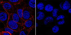
- IGF-I R/IGF1R in MCF-7 and HDLM-2 Human Cell Lines. IGF-I R/IGF1R was detected in immersion fixed MCF-7 human breast cancer cell line (positive control, left panel) and HDLM-2 human Hodgkin's lymphoma cell line (negative control, right panel) using Goat Anti-Human/Mouse IGF-I R/IGF1R Antigen Affinity-purified Polyclonal Antibody (Catalog # AF-305-NA) at 1.7 µg/mL for 3 hours at room temperature. Cells were stained using the NorthernLights™ 557-conjugated Anti-Goat IgG Secondary Antibody (red; Catalog # NL001) and counterstained with DAPI (blue). Specific staining was localized to plasma membrane. View our protocols for Fluorescent ICC Staining of Cells on Coverslips and Fluorescent ICC Staining of Non-adherent Cells.

Supportive validation
- Submitted by
- R&D Systems (provider)
- Main image
- Experimental details
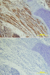
- IGF-I R/IGF1R in Mouse Embryo. IGF-I R/IGF1R was detected in immersion fixed frozen sections of mouse embryo using Goat Anti-Human IGF-I R Antigen Affinity-purified Polyclonal Antibody (Catalog # AF-305-NA) at 10 µg/mL overnight at 4 °C. Tissue was stained using the Anti-Goat HRP-DAB Cell & Tissue Staining Kit (brown; Catalog # CTS008) and counterstained with hematoxylin (blue). Lower panel shows a lack of labeling if primary antibodies are omitted and tissue is stained only with secondary antibody followed by incubation with detection reagents. View our protocol for Chromogenic IHC Staining of Frozen Tissue Sections.
- Submitted by
- R&D Systems (provider)
- Main image
- Experimental details
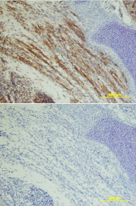
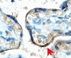
- IGF-I R/IGF1R in Human Placenta. IGF-I R/IGF1R was detected in immersion fixed paraffin-embedded sections of human placenta (chorionic villi) using 15 µg/mL Goat Anti-Human IGF-I R/IGF1R Antigen Affinity-purified Polyclonal Antibody (Catalog # AF-305-NA) overnight at 4 °C. Tissue was stained with the Anti-Goat HRP-DAB Cell & Tissue Staining Kit (brown; Catalog # CTS008) and counterstained with hematoxylin (blue). View our protocol for Chromogenic IHC Staining of Paraffin-embedded Tissue Sections.

Supportive validation
- Submitted by
- R&D Systems (provider)
- Main image
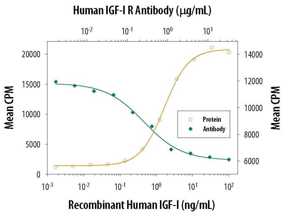
- Experimental details
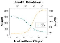
- Cell Proliferation Induced by IGF-I and Neutralization by Human IGF-I R/IGF1R Antibody. Recombinant Human IGF-I (Catalog # 291-G1) stimulates proliferation in the MCF-7 human breast cancer cell line in a dose-dependent manner (orange line). Proliferation elicited by Recombinant Human IGF-I (6 ng/mL) is neutralized (green line) by increasing concentrations of Goat Anti-Human IGF-I R/IGF1R Antigen Affinity-purified Polyclonal Antibody (Catalog # AF-305-NA). The ND50 is typically 0.5-1.5 µg/mL.