Explore
 Validate
 Learn
Learn Western blot
Antibody data
- Antibody Data
- Antigen structure
- References [3]
- Comments [0]
- Validations
- Western blot [4]
- Immunohistochemistry [1]
Submit
Validation data
Reference
Comment
Report error
- Product number
- AF8691 - Provider product page

- Provider
- R&D Systems
- Product name
- Human/Mouse/Rat p38 alpha Antibody
- Antibody type
- Polyclonal
- Description
- Antigen and protein A Affinity-purified. Detects human, mouse and rat p38 alpha . Does not detect recombinant human p38 beta , p38 gamma or p38 delta .
- Reactivity
- Human, Mouse, Rat
- Host
- Rabbit
- Conjugate
- Unconjugated
- Antigen sequence
Q16539- Isotype
- IgG
- Vial size
- 100 ug
- Concentration
- LYOPH
- Storage
- Use a manual defrost freezer and avoid repeated freeze-thaw cycles. 12 months from date of receipt, -20 to -70 °C as supplied. 1 month, 2 to 8 °C under sterile conditions after reconstitution. 6 months, -20 to -70 °C under sterile conditions after reconstitution.
Submitted references Selective p38α MAP kinase/MAPK14 inhibition in enzymatically modified LDL-stimulated human monocytes: implications for atherosclerosis.
E-selectin regulates gene expression in metastatic colorectal carcinoma cells and enhances HMGB1 release.
Interaction between the CCR5 chemokine receptors and microbial HSP70.
Cheng F, Twardowski L, Fehr S, Aner C, Schaeffeler E, Joos T, Knorpp T, Dorweiler B, Laufer S, Schwab M, Torzewski M
FASEB journal : official publication of the Federation of American Societies for Experimental Biology 2017 Feb;31(2):674-686
FASEB journal : official publication of the Federation of American Societies for Experimental Biology 2017 Feb;31(2):674-686
E-selectin regulates gene expression in metastatic colorectal carcinoma cells and enhances HMGB1 release.
Aychek T, Miller K, Sagi-Assif O, Levy-Nissenbaum O, Israeli-Amit M, Pasmanik-Chor M, Jacob-Hirsch J, Amariglio N, Rechavi G, Witz IP
International journal of cancer 2008 Oct 15;123(8):1741-50
International journal of cancer 2008 Oct 15;123(8):1741-50
Interaction between the CCR5 chemokine receptors and microbial HSP70.
Whittall T, Wang Y, Younson J, Kelly C, Bergmeier L, Peters B, Singh M, Lehner T
European journal of immunology 2006 Sep;36(9):2304-14
European journal of immunology 2006 Sep;36(9):2304-14
No comments: Submit comment
Supportive validation
- Submitted by
- R&D Systems (provider)
- Main image

- Experimental details
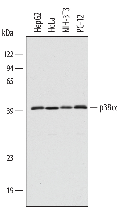
- Detection of Human, Mouse, and Rat p38 alpha by Western Blot. Western blot shows lysates of HepG2 human hepatocellular carcinoma cell line, HeLa human cervical epithelial carcinoma cell line, NIH-3T3 mouse embryonic fibroblast cell line, and PC-12 rat adrenal pheochromocytoma cell line. PVDF membrane was probed with 0.5 µg/mL Rabbit Anti-Human/Mouse/Rat p38 alpha Antigen Affinity-purified Polyclonal Antibody (Catalog # AF8691) followed by HRP-conjugated Anti-Rabbit IgG Secondary Antibody (Catalog # HAF008). A specific band for p38 alpha was detected at approximately 40 kDa (as indicated). This experiment was conducted under reducing conditions and using Immunoblot Buffer Group 1.
- Submitted by
- R&D Systems (provider)
- Main image
- Experimental details
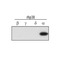
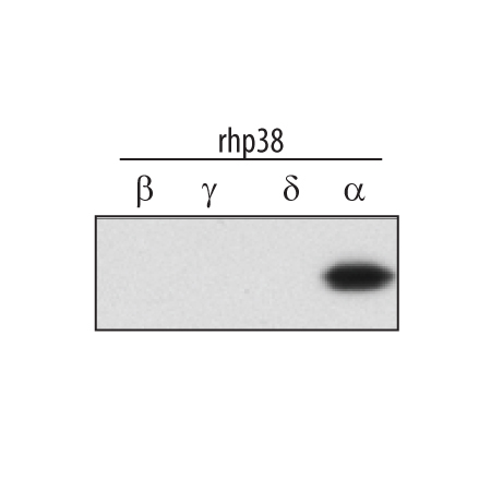
- Detection of Human p38 alpha by Western Blot. Western blot shows recombinant human p38 beta , p38 gamma , p38 delta and Recombinant Human Active p38 alpha (Catalog # 5477-KS) (2 ng/lane). PVDF membrane was probed with 0.5 µg/mL Rabbit Anti-Human/Mouse/Rat p38 alpha Antigen Affinity-purified Polyclonal Antibody (Catalog # AF8691) followed by HRP-conjugated Anti-Rabbit IgG Secondary Antibody (Catalog # HAF008). This experiment was conducted under reducing conditions and using Immunoblot Buffer Group 1.
- Submitted by
- R&D Systems (provider)
- Main image
- Experimental details
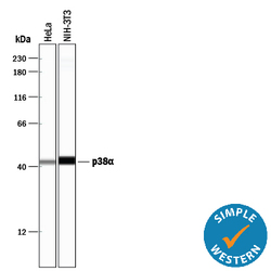
- Detection of Human and Mouse p38 alpha by Simple WesternTM. Simple Western lane view shows lysates of HeLa human cervical epithelial carcinoma cell line and NIH-3T3 mouse embryonic fibroblast cell line, loaded at 0.2 mg/mL. A specific band was detected for p38 alpha at approximately 43 kDa (as indicated) using 5 µg/mL of Rabbit Anti-Human/Mouse/Rat p38 alpha Antigen Affinity-purified Polyclonal Antibody (Catalog # AF8691). This experiment was conducted under reducing conditions and using the 12-230 kDa separation system.

- Submitted by
- R&D Systems (provider)
- Main image
- Experimental details
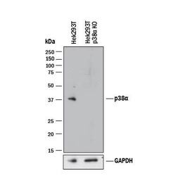
- Western Blot Shows Human p38 alpha Specificity by Using Knockout Cell Line. Western blot shows lysates of HEK293T human embryonic kidney parental cell line and p38 alpha knockout HEK293T cell line (KO). PVDF membrane was probed with 0.5 µg/mL of Rabbit Anti-Human/Mouse/Rat p38 alpha Antigen Affinity-purified Polyclonal Antibody (Catalog # AF8691) followed by HRP-conjugated Anti-Rabbit IgG Secondary Antibody (Catalog # HAF008). A specific band was detected for p38 alpha at approximately 38 kDa (as indicated) in the parental HEK293T cell line, but is not detectable in knockout HEK293T cell line. GAPDH (Catalog # AF5718) is shown as a loading control. This experiment was conducted under reducing conditions and using Immunoblot Buffer Group 1.

Supportive validation
- Submitted by
- R&D Systems (provider)
- Main image
- Experimental details
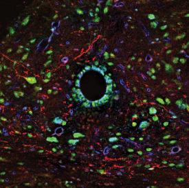
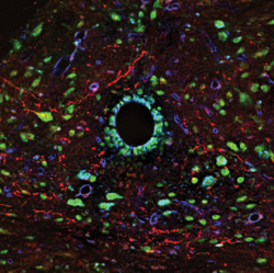
- Orexin B, p38 alpha , & Integrin beta 1 in Mouse Brainstem. Orexin B, p38 alpha , and Integrin beta 1 were detected in perfusion fixed frozen sections of mouse brainstem using Human Orexin B Monoclonal Antibody (red; Catalog # MAB734), Rabbit Anti-Human/Mouse/Rat p38 alpha Antigen Affinity-purified Polyclonal Antibody (green; Catalog # AF8691), and Mouse Integrin beta 1 Antigen Affinity-purified Polyclonal Antibody (blue; Catalog # AF2405). Tissue was incubated with primary antibodies overnight at 4 °C. Tissue was stained using NorthernLights™ 493-conjugated Anti-Rabbit IgG Secondary Antibody (green; Catalog # NL006), NorthernLights 557-conjugated Anti-Mouse IgG Secondary Antibody (red; Catalog # NL007), and NorthernLights 637-conjugated Anti-Goat IgG Secondary Antibody (blue; Catalog # NL002). The image of Integrin beta 1 is pseudo-colored for presentation. View our protocol for Fluorescent IHC Staining of Frozen Tissue Sections.