 Explore
 Validate
 Learn
Learn Western blot
Antibody data
- Antibody Data
- Antigen structure
- References [1]
- Comments [0]
- Validations
- Western blot [3]
- Immunocytochemistry [1]
- Immunohistochemistry [3]
- Other assay [1]
Submit
Validation data
Reference
Comment
Report error
- Product number
- PA5-23179 - Provider product page

- Provider
- Invitrogen Antibodies
- Product name
- c-MAF Polyclonal Antibody
- Antibody type
- Polyclonal
- Antigen
- Synthetic peptide
- Reactivity
- Human, Mouse, Rat, Bovine
- Host
- Rabbit
- Isotype
- IgG
- Vial size
- 100 µg
- Concentration
- 1.0 mg/mL
- Storage
- Store at 4°C short term. For long term storage, store at -20°C, avoiding freeze/thaw cycles.
Submitted references MicroRNA-155 contributes to plexiform neurofibroma growth downstream of MEK.
Na Y, Hall A, Choi K, Hu L, Rose J, Coover RA, Miller A, Hennigan RF, Dombi E, Kim MO, Subramanian S, Ratner N, Wu J
Oncogene 2021 Feb;40(5):951-963
Oncogene 2021 Feb;40(5):951-963
No comments: Submit comment
Supportive validation
- Submitted by
- Invitrogen Antibodies (provider)
- Main image

- Experimental details
- Western blot analysis of c-maf on Lysate from human brain in the 1) absence, 2) presence of immunizing peptide, 3) mouse brain and 4) rat brain using a c-maf polyclonal antibody (Product # PA5-23179) at 3 µg/mL.

- Submitted by
- Invitrogen Antibodies (provider)
- Main image

- Experimental details
- Western blot analysis of c-MAF in Lysate from human brain in the 1) absence, 2) presence of immunizing peptide, 3) mouse brain and 4) rat brain. Samples were incubated in c-MAF polyclonal antibody (Product # PA5-23179) using a dilution of 3 µg/mL.

- Submitted by
- Invitrogen Antibodies (provider)
- Main image

- Experimental details
- Western Blot was performed using Anti-c-MAF Polyclonal Antibody (Product # PA5-23179) and a 42 kDa band corresponding to Transcription factor MAF was observed in C2C12 cell line and increased upon differentiation to Myotubules along with uncharacterized bands around 20-35 kDa(*). Nuclear enriched extracts (30 µg lysate) of C2C12 (Lane 1) and C2C12 differentiated to Myotubules (Lane 2) were electrophoresed using NuPAGE™ 4-12% Bis-Tris Protein Gel (Product # NP0321BOX). Resolved proteins were then transferred onto a nitrocellulose membrane (Product # LC2001) by iBlot® 2 Dry Blotting System (Product # IB21001). The blot was probed with the primary antibody at a concentration of 1 µg/mL and detected by chemiluminescence with Goat anti-Rabbit IgG (H+L) Superclonal™ Recombinant Secondary Antibody, HRP (Product # A27036, 1:4000 dilution using the iBright FL 1000 (Product # A32752). Chemiluminescent detection was performed using Novex® ECL Chemiluminescent Substrate Reagent Kit (Product # WP20005).

Supportive validation
- Submitted by
- Invitrogen Antibodies (provider)
- Main image

- Experimental details
- Immunofluorescent analysis of cMAF (green) in RPMI 8226 and Daudi cells. Cells were immersion fixed in 4% PFA and permeabilized in 0.5% Triton X-100. Cells were stained with cMAF polyclonal antibody (Product # PA5-23179) at a dilution of 1:500 overnight at 4 degrees, washed in PBS followed by incubation with an Alexa 488 donkey anti-rabbit secondary antibody at a dilution of 1:100 for one hour at room temperature. Data courtesy of the Antibody Data Exchange Program.

Supportive validation
- Submitted by
- Invitrogen Antibodies (provider)
- Main image

- Experimental details
- Immunohistochemical analysis of c-MAF in formalin fixed paraffin embedded (FFPE) tissue section of human liver cancer. Samples were incubated in c-MAF polyclonal antibody (Product # PA5-23179) using a dilution of 1:500 followed by HRP conjugated anti-rabbit secondary antibody with DAB reagent. The sections were further counterstained with hematoxylin for labeling cellular nuclei. This c-MAF antibody showed a very strong nuclear and moderate to strong staining in the endothelial cells (blood vessels) and in a subset of apparently infiltrating inflammatory cells. Weak to moderate cytoplasmic and nuclear staining was observed in liver cancer cells and the cells of tumor stroma including fibroblasts.

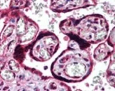
- Submitted by
- Invitrogen Antibodies (provider)
- Main image

- Experimental details
- Immunohistochemical analysis of c-MAF in Human placenta. Samples were incubated in c-MAF polyclonal antibody (Product # PA5-23179) using a dilution of 10 µg/mL.
- Submitted by
- Invitrogen Antibodies (provider)
- Main image
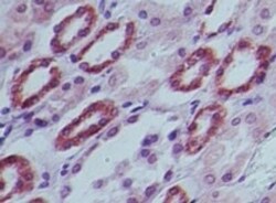
- Experimental details
- Immunohistochemical analysis of c-MAF in Human kidney. Samples were incubated in c-MAF polyclonal antibody (Product # PA5-23179) using a dilution of 10 µg/mL.

Supportive validation
- Submitted by
- Invitrogen Antibodies (provider)
- Main image
- Experimental details
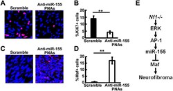
- Figure 7. Anti-miR-155-PNAs effectively inhibits cell proliferation and increased target protein expression. A . Representative immunofluorescence pictures of Ki67+ staining (Red) in scramble (left) and anti-miR-155-PNA (right) treated mouse plexiform neurofibromas. DAPI (blue) was used for nuclei labelling. B. Quantification of Ki67+% cells in scramble (black bar, n=3) and anti-miR-155-PNA (white bar, n=4) treated mouse plexiform neurofibromas. C . Representative immunofluorescence pictures of MAF+ staining (Red) in scramble (left) and anti-miR-155-PNA (right) treated mouse plexiform neurofibromas. DAPI (blue) was used for nuclei labelling. D. Quantification of MAF+% cells in scramble (black bar, n=3) and anti-miR-155-PNA (white bar, n=4) treated mouse plexiform neurofibromas. E. Schematic showing a model of PNF formation: Loss of Nf1 in SC/SCP elevated MEK/ERK levels to activate AP-1. AP-1 binds to miR-155, which in turn regulates one of its main targets, Maf, to contribute to PNF formation.
