Antibody data
- Antibody Data
- Antigen structure
- References [0]
- Comments [0]
- Validations
- Western blot [3]
- Immunocytochemistry [1]
- Immunohistochemistry [1]
Submit
Validation data
Reference
Comment
Report error
- Product number
- GTX48691 - Provider product page

- Provider
- GeneTex
- Proper citation
- GeneTex Cat#GTX48691, RRID:AB_11176335
- Product name
- Jagged 1 antibody
- Antibody type
- Polyclonal
- Reactivity
- Human, Mouse
- Host
- Rabbit
No comments: Submit comment
Supportive validation
- Submitted by
- GeneTex (provider)
- Main image

- Experimental details
- Western blot using GeneTex's Protein A purified anti-Jagged-1 antibody shows detection of Jagged-1 protein in various whole cell lysates: human brain (lane 1), human kidney (lane 2), human liver (lane 3), and mouse liver (lane 5). Lane 4 contained sample buffer only. The band at ~134 kDa in lane 5 is believed to be Jagged-1 precursor. The identity of minor reactive bands is unknown. Each lane contains approximately 20 µg of lysate. Primary antibody was used at a 1:500 dilution. The membrane was washed and reacted with a 1:5,000 dilution of HRP conjugated goat anti-Rabbit IgG. Exposure time was 1 min. Predicted molecular weight is 134 kDa.
- Validation comment
- WB
- Submitted by
- GeneTex (provider)
- Main image

- Experimental details
- Western blot using GeneTex's Protein A purified anti-Jagged-1 antibody shows detection of Jagged-1 protein in various whole cell lysates: human brain (lane 1), human kidney (lane 2), human liver (lane 3), and mouse liver (lane 5). Lane 4 contained sample buffer only. The band at ~134 kDa in lane 5 is believed to be Jagged-1 precursor. The identity of minor reactive bands is unknown. Each lane contains approximately 20 ?g of lysate. Primary antibody was used at a 1:500 dilution. The membrane was washed and reacted with a 1:5,000 dilution of HRP conjugated goat anti-Rabbit IgG. Exposure time was 1 min. Predicted molecular weight is 134 kDa.
- Submitted by
- GeneTex (provider)
- Main image

- Experimental details
- WB analysis of mouse liver using GTX48691Jagged 1 antibody at 1:500.
Supportive validation
- Submitted by
- GeneTex (provider)
- Main image

- Experimental details
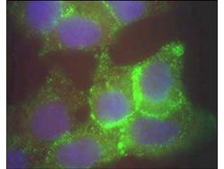
- Immunofluorescence Microscopy using GeneTex's Protein A purified anti-Jagged-1 antibody of human corneal epithelial cells. Primary antibody was used at a 1:500 dilution. The Jagged1 Green staining) is localized to the cytoplasm and is consistent with reports in the literature. The nucleus is stained with Bis benzimide (blue)
Supportive validation
- Submitted by
- GeneTex (provider)
- Main image

- Experimental details
- Immunohistochemical staining of human cervical cancer tissue (40X magnification) using GeneTex's Protein A purified anti-Jagged-1 antibody. Tissue was fixed with formalin and embedded in paraffin. Hematoxylin was used to counter-stain cells. A 1:100 dilution of primary antibody was used.
 Explore
 Validate
 Learn
Learn Western blot
 ELISA
ELISA