Explore
 Validate
 Learn
Learn Western blot
Antibody data
- Antibody Data
- Antigen structure
- References [1]
- Comments [0]
- Validations
- Western blot [3]
- Immunohistochemistry [1]
- Other assay [3]
Submit
Validation data
Reference
Comment
Report error
- Product number
- PA5-46970 - Provider product page

- Provider
- Invitrogen Antibodies
- Product name
- Jagged1 Polyclonal Antibody
- Antibody type
- Polyclonal
- Antigen
- Recombinant full-length protein
- Description
- In direct ELISAs, approximately 20% cross-reactivity with recombinant rat Jagged 1 is observed and less than 1% cross-reactivity with recombinant human Jagged 2 is observed.
- Concentration
- 0.2 mg/mL
Submitted references Notch1 signaling determines the plasticity and function of fibroblasts in diabetic wounds.
Shao H, Li Y, Pastar I, Xiao M, Prokupets R, Liu S, Yu K, Vazquez-Padron RI, Tomic-Canic M, Velazquez OC, Liu ZJ
Life science alliance 2020 Dec;3(12)
Life science alliance 2020 Dec;3(12)
No comments: Submit comment
Supportive validation
- Submitted by
- Invitrogen Antibodies (provider)
- Main image
- Experimental details
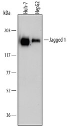
- Western blot analysis from lysates of Huh-7 human hepatoma cell line and HepG2 human hepatocellular carcinoma cell line. PVDF Membrane was probed with 1 µg/mL of Goat Anti-human Jagged 1 Antigen Affinity-purified Polyclonal Antibody (Product # PA5-46970) followed by HRP-conjugated Anti-Goat IgG Secondary Antibody. A specific band was detected for Jagged 1 at approximately 180 kDa (as indicated). This experiment was conducted under reducing conditions.

- Submitted by
- Invitrogen Antibodies (provider)
- Main image

- Experimental details
- Western blot analysis of Jagged1 in Huh‚7 human hepatoma cell line and HepG2 human hepatocellular carcinoma cell line. Samples were incubated in Jagged1 polyclonal antibody (Product # PA5-46970) using a dilution of 1 µg/mL followed by a HRP-conjugated Anti-Goat IgG secondary antibody. A specific band was detected for Jagged 1 at approximately 180 kDa (as indicated). This experiment was conducted under reducing conditions.

- Submitted by
- Invitrogen Antibodies (provider)
- Main image

- Experimental details
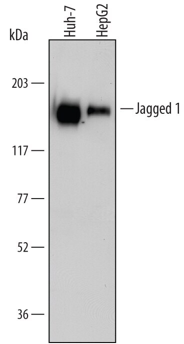
- Western blot analysis of Jagged1 in Huh‚7 human hepatoma cell line and HepG2 human hepatocellular carcinoma cell line. Samples were incubated in Jagged1 polyclonal antibody (Product # PA5-46970) using a dilution of 1 µg/mL followed by a HRP-conjugated Anti-Goat IgG secondary antibody. A specific band was detected for Jagged 1 at approximately 180 kDa (as indicated). This experiment was conducted under reducing conditions.
Supportive validation
- Submitted by
- Invitrogen Antibodies (provider)
- Main image

- Experimental details
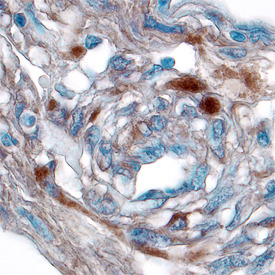
- Immunohistochemical analysis of Jagged1 in immersion fixed paraffin-embedded sections of human kidney cancer tissue. Samples were incubated in Jagged1 polyclonal antibody (Product # PA5-46970) using a dilution of 15 µg/mL overnight at 4 °C. Tissue was stained using the Anti-Goat HRP-DAB Cell & Tissue Staining Kit (brown) and counterstained with hematoxylin (blue).
Supportive validation
- Submitted by
- Invitrogen Antibodies (provider)
- Main image
- Experimental details
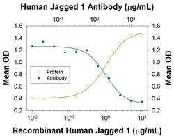
- Neutralization antibody testing demonstrates the specificity of an antibody through a correlation between antibody binding and the activity of the target. Neutralization of Jagged1 is shown by the decrease in mean OD (measure of alkaline phosphatase production by C3H10T1/2 mouse embryonic fibroblast cell line) with increasing concentrations of Jagged1 Polyclonal Antibody (PA5-46970).

- Submitted by
- Invitrogen Antibodies (provider)
- Main image

- Experimental details
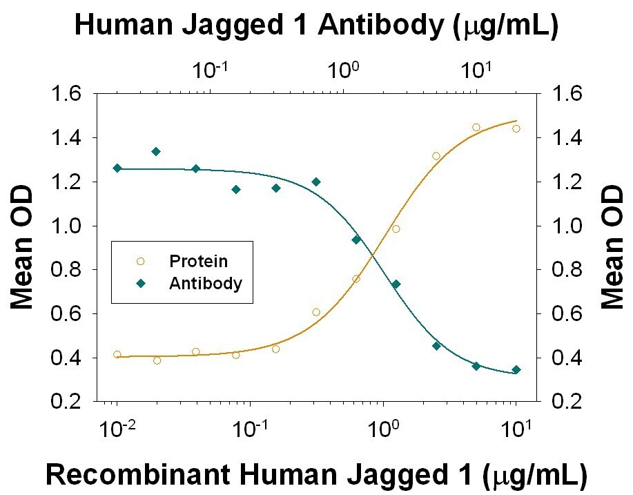
- Neutralization of Jagged1 in C3H10T1/2 mouse embryonic fibroblast cell line. Samples were incubated in Jagged1 polyclonal antibody (Product # PA5-46970). Recombinant Human Jagged 1 Fc Chimera induces alkaline phosphatase production in the C3H10T1/2 mouse embryonic fibroblast cell line in the presence of Recombinant Human/Mouse/Rat BMP-2 in a dose-dependent manner (orange line). Alkaline phosphatase production elicited by Recombinant Human Jagged 1 Fc Chimera (5 µg/mL) is neutralized (green line) by increasing concentrations of Goat Anti-Human Jagged 1 Antibody. The ND50 is typically 1-5 µg/mL.
- Submitted by
- Invitrogen Antibodies (provider)
- Main image

- Experimental details
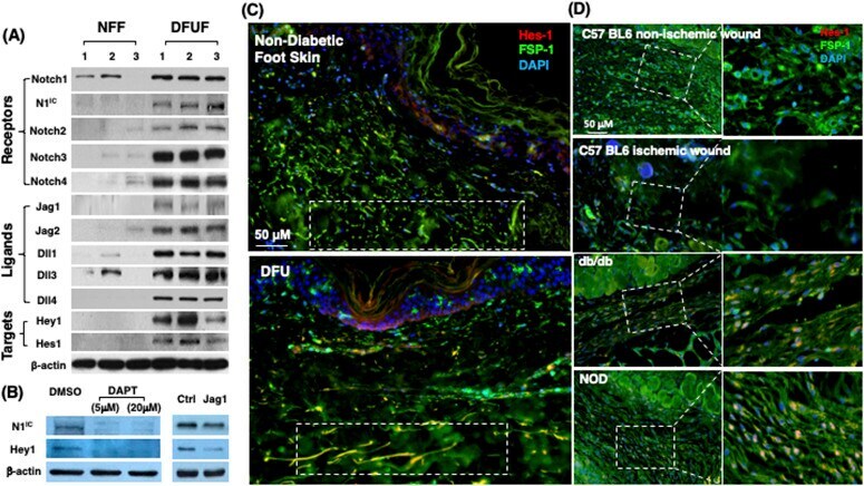
- Figure 1. Differential Notch pathway activities in fibroblasts of chronic diabetic skin wounds versus non-diabetic skin and wounds. (A) High Notch pathway activity in diabetic foot ulcer fibroblasts (DFUF) versus low Notch pathway activity in normal foot fibroblasts (NFF). Expression of Notch pathway components in three DFUF and three NFF were assessed by immunoblot. beta-actin was used as a loading control. The band of each molecule is shown. (B) Inhibition of the Notch pathway activity, reflected by decreased levels of N1 IC and Hey-1, in DFUF by DAPT and Jag 1 neutralizing Ab. Compared with DAPT, Jag 1 neutralizing Ab only achieved a partial inhibition. (C) Representative immunostaining images show that fibroblasts (green) express higher levels of Hes-1 (red) in skin at the edge of diabetic foot ulcer tissue than that in non-diabetic foot skin. Highlighted areas show fibroblasts in reticular layers. (D) Representative immunostaining images show that fibroblasts (green) express higher levels of Hes-1 (red) in wounds of diabetic mice (db/db and NOD) but not in non-diabetic acute wound and ischemic chronic wounds in C57 BL6 mice. Wound tissues were harvested at day 7. Highlighted areas show fibroblasts in granulation tissues.