Explore
 Validate
 Learn
Learn Western blot
 ELISA
ELISAAntibody data
- Antibody Data
- Antigen structure
- References [0]
- Comments [0]
- Validations
- Western blot [2]
- Immunohistochemistry [25]
Submit
Validation data
Reference
Comment
Report error
- Product number
- STJ95727 - Provider product page

- Provider
- St John's Laboratory
- Product name
- Anti-SOCS1 antibody (Internal) (STJ95727)
- Antibody type
- Polyclonal
- Description
- Rabbit polyclonal antibody anti-Suppressor Of Cytokine Signaling 1 (Internal) is suitable for use in Immunofluorescence, Immunocytochemistry, Western Blot, Immunohistochemistry and ELISA research applications.
- Reactivity
- Human, Mouse, Rat
- Host
- Rabbit
- Conjugate
- Unconjugated
- Antigen sequence
NA- Epitope
- NA
- Isotype
- IgG
- Antibody clone number
- NA
- Vial size
- NA
- Concentration
- NA
- Storage
- Store at-20°C for up to 1 year from the date of receipt, and avoid repeat freeze-thaw cycles.
- Handling
- NA
No comments: Submit comment
Supportive validation
Supportive validation
- Submitted by
- St John's Laboratory (provider)
- Main image
- Experimental details
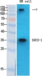
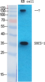
- Western blot analysis of various cells using SOCS-1 Polyclonal Antibody diluted at 1ï¼2000
- Sample type
- NA
- Validation comment
- NA
- Primary Ab dilution
- NA
- Other comments
- NA
- Secondary Ab
- NA
- Secondary Ab dilution
- NA
- Protocol
- NA
Supportive validation
- Submitted by
- St John's Laboratory (provider)
- Main image
- Experimental details
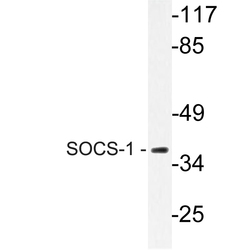
- Western blot analysis of lysate from Jurkat cells, using SOCS-1 antibody.
- Sample type
- NA
- Validation comment
- NA
- Primary Ab dilution
- NA
- Other comments
- NA
- Secondary Ab
- NA
- Secondary Ab dilution
- NA
- Protocol
- NA

Supportive validation
Supportive validation
Supportive validation
Supportive validation
Supportive validation
Supportive validation
Supportive validation
Supportive validation
Supportive validation
Supportive validation
Supportive validation
Supportive validation
Supportive validation
Supportive validation
Supportive validation
Supportive validation
Supportive validation
Supportive validation
Supportive validation
Supportive validation
Supportive validation
Supportive validation
Supportive validation
Supportive validation
Supportive validation
- Submitted by
- St John's Laboratory (provider)
- Main image
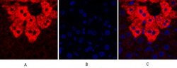
- Experimental details
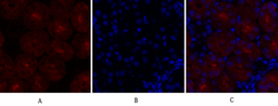
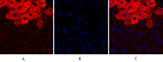
- Immunofluorescence analysis of mouse-liver tissue. 1, SOCS-1 Polyclonal Antibody (red) was diluted at 1:200 (4°C, overnight). 2, Cy3 labled Secondary antibody was diluted at 1:300 (room temperature, 50min).3, Picture B: DAPI (blue) 10min. Picture A:Target. Picture B: DAPI. Picture C: merge of A+B
- Sample type
- NA
- Validation comment
- NA
- Primary Ab dilution
- NA
- Other comments
- NA
- Secondary Ab
- NA
- Secondary Ab dilution
- NA
- Protocol
- NA
Supportive validation
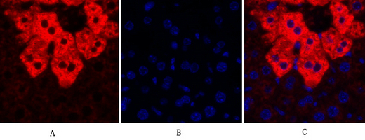
- Submitted by
- St John's Laboratory (provider)
- Main image
- Experimental details
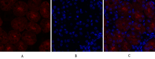
- Immunofluorescence analysis of mouse-kidney tissue. 1, SOCS-1 Polyclonal Antibody (red) was diluted at 1:200 (4°C, overnight). 2, Cy3 labled Secondary antibody was diluted at 1:300 (room temperature, 50min).3, Picture B: DAPI (blue) 10min. Picture A:Target. Picture B: DAPI. Picture C: merge of A+B
- Sample type
- NA
- Validation comment
- NA
- Primary Ab dilution
- NA
- Other comments
- NA
- Secondary Ab
- NA
- Secondary Ab dilution
- NA
- Protocol
- NA
Supportive validation
- Submitted by
- St John's Laboratory (provider)
- Main image
- Experimental details
- Immunofluorescence analysis of mouse-kidney tissue. 1, SOCS-1 Polyclonal Antibody (red) was diluted at 1:200 (4°C, overnight). 2, Cy3 labled Secondary antibody was diluted at 1:300 (room temperature, 50min).3, Picture B: DAPI (blue) 10min. Picture A:Target. Picture B: DAPI. Picture C: merge of A+B
- Sample type
- NA
- Validation comment
- NA
- Primary Ab dilution
- NA
- Other comments
- NA
- Secondary Ab
- NA
- Secondary Ab dilution
- NA
- Protocol
- NA
Supportive validation
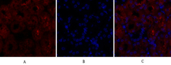
- Submitted by
- St John's Laboratory (provider)
- Main image
- Experimental details
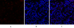
- Immunofluorescence analysis of mouse-lung tissue. 1, SOCS-1 Polyclonal Antibody (red) was diluted at 1:200 (4°C, overnight). 2, Cy3 labled Secondary antibody was diluted at 1:300 (room temperature, 50min).3, Picture B: DAPI (blue) 10min. Picture A:Target. Picture B: DAPI. Picture C: merge of A+B
- Sample type
- NA
- Validation comment
- NA
- Primary Ab dilution
- NA
- Other comments
- NA
- Secondary Ab
- NA
- Secondary Ab dilution
- NA
- Protocol
- NA
Supportive validation
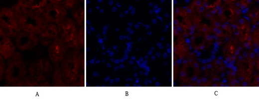
- Submitted by
- St John's Laboratory (provider)
- Main image
- Experimental details
- Immunofluorescence analysis of mouse-lung tissue. 1, SOCS-1 Polyclonal Antibody (red) was diluted at 1:200 (4°C, overnight). 2, Cy3 labled Secondary antibody was diluted at 1:300 (room temperature, 50min).3, Picture B: DAPI (blue) 10min. Picture A:Target. Picture B: DAPI. Picture C: merge of A+B
- Sample type
- NA
- Validation comment
- NA
- Primary Ab dilution
- NA
- Other comments
- NA
- Secondary Ab
- NA
- Secondary Ab dilution
- NA
- Protocol
- NA

Supportive validation
- Submitted by
- St John's Laboratory (provider)
- Main image
- Experimental details
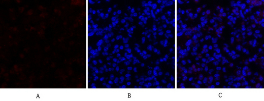
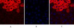
- Immunofluorescence analysis of mouse-liver tissue. 1, SOCS-1 Polyclonal Antibody (red) was diluted at 1:200 (4°C, overnight). 2, Cy3 labled Secondary antibody was diluted at 1:300 (room temperature, 50min).3, Picture B: DAPI (blue) 10min. Picture A:Target. Picture B: DAPI. Picture C: merge of A+B
- Sample type
- NA
- Validation comment
- NA
- Primary Ab dilution
- NA
- Other comments
- NA
- Secondary Ab
- NA
- Secondary Ab dilution
- NA
- Protocol
- NA
Supportive validation
- Submitted by
- St John's Laboratory (provider)
- Main image

- Experimental details
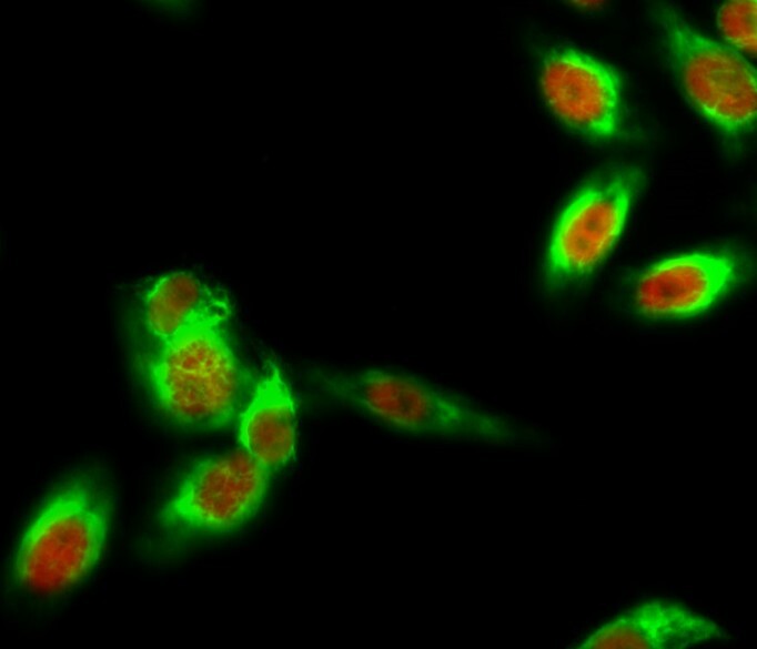
- Immunofluorescence analysis of Hela cell. 1, SOCS-1 Polyclonal Antibody (red) was diluted at 1:200 (4°C overnight). Beta-Tubulin monoclonal antibody (5G3) (green) was diluted at 1:200 (4°C overnight). 2, Goat Anti Rabbit Alexa Fluor 594 Catalog: (NA was diluted at 1:1000 (room temperature, 50min). Goat Anti Mouse Alexa Fluor 488 Catalog: (NA was diluted at 1:1000 (room temperature, 50min).
- Sample type
- NA
- Validation comment
- NA
- Primary Ab dilution
- NA
- Other comments
- NA
- Secondary Ab
- NA
- Secondary Ab dilution
- NA
- Protocol
- NA
Supportive validation
- Submitted by
- St John's Laboratory (provider)
- Main image

- Experimental details
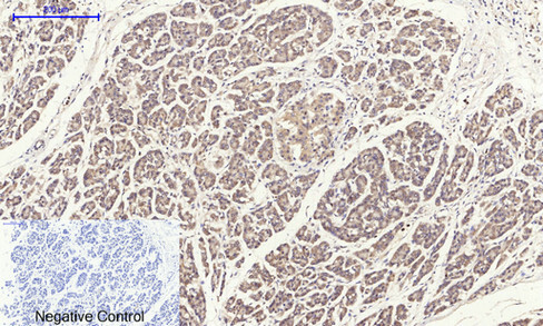
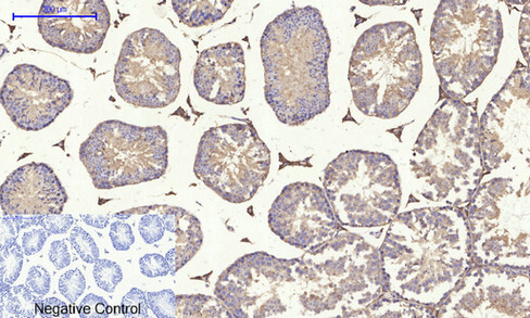
- Immunohistochemical analysis of paraffin-embedded Human-stomach-cancer tissue. 1, SOCS-1 Polyclonal Antibody was diluted at 1:200 (4°C, overnight). 2, Sodium citrate pH 6.0 was used for antibody retrieval (>98°C, 20min). 3, Secondary antibody was diluted at 1:200 (room tempeRature, 30min). Negative control was used by secondary antibody only.
- Sample type
- NA
- Validation comment
- NA
- Primary Ab dilution
- NA
- Other comments
- NA
- Secondary Ab
- NA
- Secondary Ab dilution
- NA
- Protocol
- NA
Supportive validation
- Submitted by
- St John's Laboratory (provider)
- Main image
- Experimental details
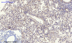
- Immunohistochemical analysis of paraffin-embedded Mouse-lung tissue. 1, SOCS-1 Polyclonal Antibody was diluted at 1:200 (4°C, overnight). 2, Sodium citrate pH 6.0 was used for antibody retrieval (>98°C, 20min). 3, Secondary antibody was diluted at 1:200 (room tempeRature, 30min). Negative control was used by secondary antibody only.
- Sample type
- NA
- Validation comment
- NA
- Primary Ab dilution
- NA
- Other comments
- NA
- Secondary Ab
- NA
- Secondary Ab dilution
- NA
- Protocol
- NA
Supportive validation
- Submitted by
- St John's Laboratory (provider)
- Main image

- Experimental details
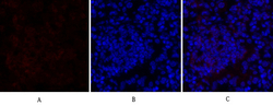
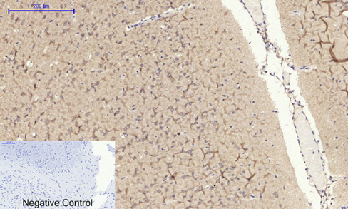
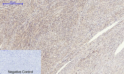
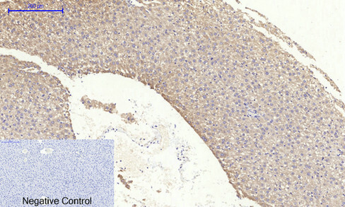
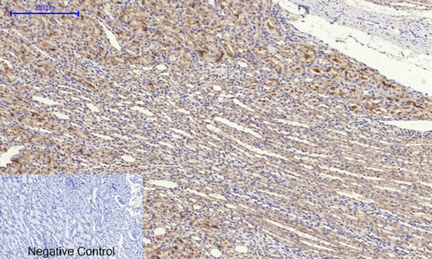
- Immunohistochemical analysis of paraffin-embedded Rat-brain tissue. 1, SOCS-1 Polyclonal Antibody was diluted at 1:200 (4°C, overnight). 2, Sodium citrate pH 6.0 was used for antibody retrieval (>98°C, 20min). 3, Secondary antibody was diluted at 1:200 (room tempeRature, 30min). Negative control was used by secondary antibody only.
- Sample type
- NA
- Validation comment
- NA
- Primary Ab dilution
- NA
- Other comments
- NA
- Secondary Ab
- NA
- Secondary Ab dilution
- NA
- Protocol
- NA
Supportive validation
- Submitted by
- St John's Laboratory (provider)
- Main image

- Experimental details
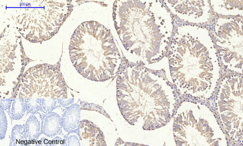
- Immunohistochemical analysis of paraffin-embedded Mouse-testis tissue. 1, SOCS-1 Polyclonal Antibody was diluted at 1:200 (4°C, overnight). 2, Sodium citrate pH 6.0 was used for antibody retrieval (>98°C, 20min). 3, Secondary antibody was diluted at 1:200 (room tempeRature, 30min). Negative control was used by secondary antibody only.
- Sample type
- NA
- Validation comment
- NA
- Primary Ab dilution
- NA
- Other comments
- NA
- Secondary Ab
- NA
- Secondary Ab dilution
- NA
- Protocol
- NA
Supportive validation
- Submitted by
- St John's Laboratory (provider)
- Main image
- Experimental details
- Immunohistochemical analysis of paraffin-embedded Human-uterus tissue. 1, SOCS-1 Polyclonal Antibody was diluted at 1:200 (4°C, overnight). 2, Sodium citrate pH 6.0 was used for antibody retrieval (>98°C, 20min). 3, Secondary antibody was diluted at 1:200 (room tempeRature, 30min). Negative control was used by secondary antibody only.
- Sample type
- NA
- Validation comment
- NA
- Primary Ab dilution
- NA
- Other comments
- NA
- Secondary Ab
- NA
- Secondary Ab dilution
- NA
- Protocol
- NA
Supportive validation
- Submitted by
- St John's Laboratory (provider)
- Main image
- Experimental details
- Immunohistochemical analysis of paraffin-embedded Rat-liver tissue. 1, SOCS-1 Polyclonal Antibody was diluted at 1:200 (4°C, overnight). 2, Sodium citrate pH 6.0 was used for antibody retrieval (>98°C, 20min). 3, Secondary antibody was diluted at 1:200 (room tempeRature, 30min). Negative control was used by secondary antibody only.
- Sample type
- NA
- Validation comment
- NA
- Primary Ab dilution
- NA
- Other comments
- NA
- Secondary Ab
- NA
- Secondary Ab dilution
- NA
- Protocol
- NA
Supportive validation
- Submitted by
- St John's Laboratory (provider)
- Main image

- Experimental details
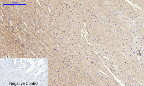
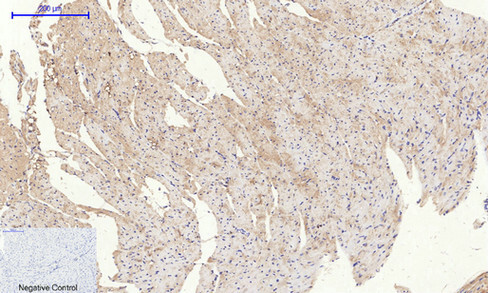
- Immunohistochemical analysis of paraffin-embedded Rat-heart tissue. 1, SOCS-1 Polyclonal Antibody was diluted at 1:200 (4°C, overnight). 2, Sodium citrate pH 6.0 was used for antibody retrieval (>98°C, 20min). 3, Secondary antibody was diluted at 1:200 (room tempeRature, 30min). Negative control was used by secondary antibody only.
- Sample type
- NA
- Validation comment
- NA
- Primary Ab dilution
- NA
- Other comments
- NA
- Secondary Ab
- NA
- Secondary Ab dilution
- NA
- Protocol
- NA
Supportive validation
- Submitted by
- St John's Laboratory (provider)
- Main image
- Experimental details
- Immunohistochemical analysis of paraffin-embedded Human-uterus-cancer tissue. 1, SOCS-1 Polyclonal Antibody was diluted at 1:200 (4°C, overnight). 2, Sodium citrate pH 6.0 was used for antibody retrieval (>98°C, 20min). 3, Secondary antibody was diluted at 1:200 (room tempeRature, 30min). Negative control was used by secondary antibody only.
- Sample type
- NA
- Validation comment
- NA
- Primary Ab dilution
- NA
- Other comments
- NA
- Secondary Ab
- NA
- Secondary Ab dilution
- NA
- Protocol
- NA

Supportive validation
- Submitted by
- St John's Laboratory (provider)
- Main image
- Experimental details
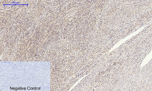
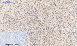
- Immunohistochemical analysis of paraffin-embedded Rat-testis tissue. 1, SOCS-1 Polyclonal Antibody was diluted at 1:200 (4°C, overnight). 2, Sodium citrate pH 6.0 was used for antibody retrieval (>98°C, 20min). 3, Secondary antibody was diluted at 1:200 (room tempeRature, 30min). Negative control was used by secondary antibody only.
- Sample type
- NA
- Validation comment
- NA
- Primary Ab dilution
- NA
- Other comments
- NA
- Secondary Ab
- NA
- Secondary Ab dilution
- NA
- Protocol
- NA
Supportive validation
- Submitted by
- St John's Laboratory (provider)
- Main image
- Experimental details
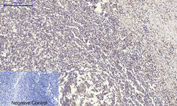
- Immunohistochemical analysis of paraffin-embedded Human-Tonsil tissue. 1, SOCS-1 Polyclonal Antibody was diluted at 1:200 (4°C, overnight). 2, Sodium citrate pH 6.0 was used for antibody retrieval (>98°C, 20min). 3, Secondary antibody was diluted at 1:200 (room tempeRature, 30min). Negative control was used by secondary antibody only.
- Sample type
- NA
- Validation comment
- NA
- Primary Ab dilution
- NA
- Other comments
- NA
- Secondary Ab
- NA
- Secondary Ab dilution
- NA
- Protocol
- NA

Supportive validation
- Submitted by
- St John's Laboratory (provider)
- Main image

- Experimental details
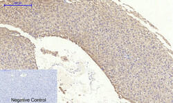
- Immunohistochemical analysis of paraffin-embedded Rat-kidney tissue. 1, SOCS-1 Polyclonal Antibody was diluted at 1:200 (4°C, overnight). 2, Sodium citrate pH 6.0 was used for antibody retrieval (>98°C, 20min). 3, Secondary antibody was diluted at 1:200 (room tempeRature, 30min). Negative control was used by secondary antibody only.
- Sample type
- NA
- Validation comment
- NA
- Primary Ab dilution
- NA
- Other comments
- NA
- Secondary Ab
- NA
- Secondary Ab dilution
- NA
- Protocol
- NA
Supportive validation
- Submitted by
- St John's Laboratory (provider)
- Main image

- Experimental details
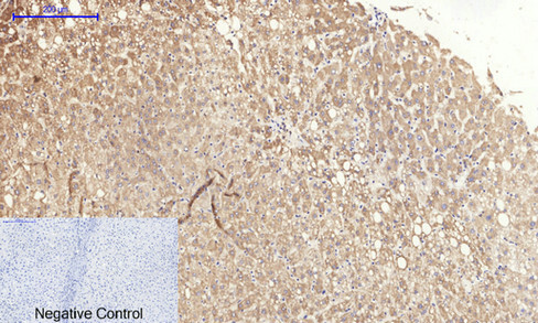
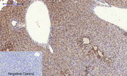
- Immunohistochemical analysis of paraffin-embedded Human-liver tissue. 1, SOCS-1 Polyclonal Antibody was diluted at 1:200 (4°C, overnight). 2, Sodium citrate pH 6.0 was used for antibody retrieval (>98°C, 20min). 3, Secondary antibody was diluted at 1:200 (room tempeRature, 30min). Negative control was used by secondary antibody only.
- Sample type
- NA
- Validation comment
- NA
- Primary Ab dilution
- NA
- Other comments
- NA
- Secondary Ab
- NA
- Secondary Ab dilution
- NA
- Protocol
- NA
Supportive validation
- Submitted by
- St John's Laboratory (provider)
- Main image

- Experimental details
- Immunohistochemical analysis of paraffin-embedded Mouse-heart tissue. 1, SOCS-1 Polyclonal Antibody was diluted at 1:200 (4°C, overnight). 2, Sodium citrate pH 6.0 was used for antibody retrieval (>98°C, 20min). 3, Secondary antibody was diluted at 1:200 (room tempeRature, 30min). Negative control was used by secondary antibody only.
- Sample type
- NA
- Validation comment
- NA
- Primary Ab dilution
- NA
- Other comments
- NA
- Secondary Ab
- NA
- Secondary Ab dilution
- NA
- Protocol
- NA
Supportive validation
- Submitted by
- St John's Laboratory (provider)
- Main image

- Experimental details
- Immunohistochemical analysis of paraffin-embedded Human-liver-cancer tissue. 1, SOCS-1 Polyclonal Antibody was diluted at 1:200 (4°C, overnight). 2, Sodium citrate pH 6.0 was used for antibody retrieval (>98°C, 20min). 3, Secondary antibody was diluted at 1:200 (room tempeRature, 30min). Negative control was used by secondary antibody only.
- Sample type
- NA
- Validation comment
- NA
- Primary Ab dilution
- NA
- Other comments
- NA
- Secondary Ab
- NA
- Secondary Ab dilution
- NA
- Protocol
- NA

Supportive validation
- Submitted by
- St John's Laboratory (provider)
- Main image
- Experimental details
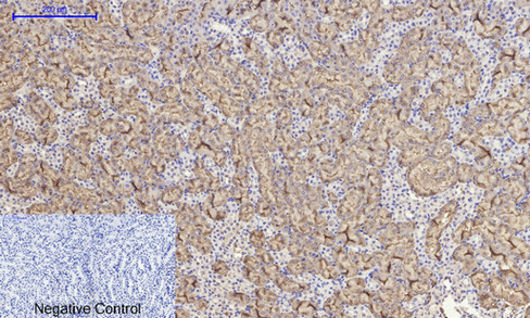
- Immunohistochemical analysis of paraffin-embedded Mouse-liver tissue. 1, SOCS-1 Polyclonal Antibody was diluted at 1:200 (4°C, overnight). 2, Sodium citrate pH 6.0 was used for antibody retrieval (>98°C, 20min). 3, Secondary antibody was diluted at 1:200 (room tempeRature, 30min). Negative control was used by secondary antibody only.
- Sample type
- NA
- Validation comment
- NA
- Primary Ab dilution
- NA
- Other comments
- NA
- Secondary Ab
- NA
- Secondary Ab dilution
- NA
- Protocol
- NA

Supportive validation
- Submitted by
- St John's Laboratory (provider)
- Main image
- Experimental details
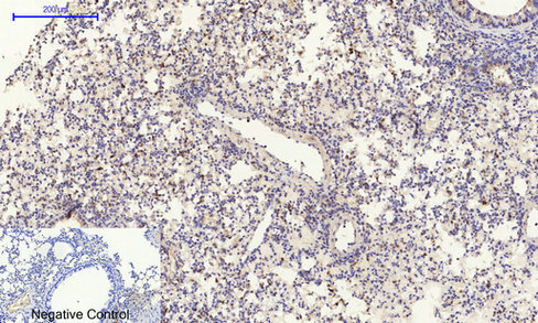
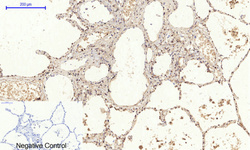
- Immunohistochemical analysis of paraffin-embedded Human-lung tissue. 1, SOCS-1 Polyclonal Antibody was diluted at 1:200 (4°C, overnight). 2, Sodium citrate pH 6.0 was used for antibody retrieval (>98°C, 20min). 3, Secondary antibody was diluted at 1:200 (room tempeRature, 30min). Negative control was used by secondary antibody only.
- Sample type
- NA
- Validation comment
- NA
- Primary Ab dilution
- NA
- Other comments
- NA
- Secondary Ab
- NA
- Secondary Ab dilution
- NA
- Protocol
- NA

Supportive validation
- Submitted by
- St John's Laboratory (provider)
- Main image

- Experimental details
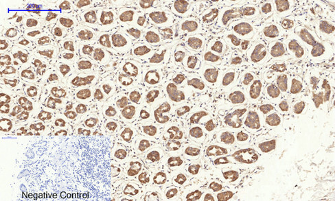
- Immunohistochemical analysis of paraffin-embedded Mouse-kidney tissue. 1, SOCS-1 Polyclonal Antibody was diluted at 1:200 (4°C, overnight). 2, Sodium citrate pH 6.0 was used for antibody retrieval (>98°C, 20min). 3, Secondary antibody was diluted at 1:200 (room tempeRature, 30min). Negative control was used by secondary antibody only.
- Sample type
- NA
- Validation comment
- NA
- Primary Ab dilution
- NA
- Other comments
- NA
- Secondary Ab
- NA
- Secondary Ab dilution
- NA
- Protocol
- NA
Supportive validation
- Submitted by
- St John's Laboratory (provider)
- Main image

- Experimental details
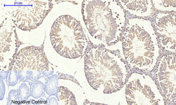
- Immunohistochemical analysis of paraffin-embedded Human-stomach tissue. 1, SOCS-1 Polyclonal Antibody was diluted at 1:200 (4°C, overnight). 2, Sodium citrate pH 6.0 was used for antibody retrieval (>98°C, 20min). 3, Secondary antibody was diluted at 1:200 (room tempeRature, 30min). Negative control was used by secondary antibody only.
- Sample type
- NA
- Validation comment
- NA
- Primary Ab dilution
- NA
- Other comments
- NA
- Secondary Ab
- NA
- Secondary Ab dilution
- NA
- Protocol
- NA