 Explore
 Validate
 Learn
Learn Western blot
Antibody data
- Antibody Data
- Antigen structure
- References [1]
- Comments [0]
- Validations
- Western blot [3]
- Immunohistochemistry [2]
- Other assay [1]
Submit
Validation data
Reference
Comment
Report error
- Product number
- PA5-70360 - Provider product page

- Provider
- Invitrogen Antibodies
- Product name
- PKA alpha Polyclonal Antibody
- Antibody type
- Polyclonal
- Antigen
- Synthetic peptide
- Description
- This target displays homology in the following species: Cow: 100%; Dog: 100%; Guinea Pig: 100%; Horse: 100%; Human: 100%; Mouse: 100%; Rabbit: 100%; Rat: 100%; Sheep: 100%; Zebrafish: 100%
- Reactivity
- Human, Mouse
- Host
- Rabbit
- Isotype
- IgG
- Vial size
- 100 µL
- Concentration
- 0.5 mg/mL
- Storage
- -20° C, Avoid Freeze/Thaw Cycles
Submitted references Functional Heterogeneity of Protein Kinase A Activation in Multipotent Stromal Cells.
Tyurin-Kuzmin PA, Karagyaur MN, Kulebyakin KY, Dyikanov DT, Chechekhin VI, Ivanova AM, Skryabina MN, Arbatskiy MS, Sysoeva VY, Kalinina NI, Tkachuk VA
International journal of molecular sciences 2020 Jun 22;21(12)
International journal of molecular sciences 2020 Jun 22;21(12)
No comments: Submit comment
Supportive validation
- Submitted by
- Invitrogen Antibodies (provider)
- Main image

- Experimental details
- Western blot analysis of PKA-alpha on mouse heart tissue lysate. The sample was probed with a PKA-alpha polyclonal antibody (Product # PA5-70360) using a primary antibody dilution of 1.0 µg/mL.

- Submitted by
- Invitrogen Antibodies (provider)
- Main image

- Experimental details
- Western blot analysis of PKA-alpha on human placenta tissue lysate. The sample was probed with a PKA-alpha polyclonal antibody (Product # PA5-70360) using a primary antibody dilution of 0.2-1.0 µg/mL.

- Submitted by
- Invitrogen Antibodies (provider)
- Main image

- Experimental details
- Western blot was performed using anti-PKA alpha Polyclonal Antibody (Product # PA5-70360) and a 40 kDa band corresponding to PRKACA was observed across cell lines and tissues tested along with an uncharacterized band at ~20, 100 kDa. Whole cell extracts (30 µg lysate) of NIH:OVCAR-3 (Lane 1), A 549 (Lane 2), Jurkat (Lane 3), NIH: 3T3 (Lane 4), C2C12 (Lane 5), HeLa (Lane 6), Mouse lung (Lane 7), Rat lung (Lane 8), Mouse kidney (Lane 9), Rat kidney (Lane 10) were electrophoresed using NuPAGE® 4-12 % Bis-Tris gel (Product # NP0321BOX). Resolved proteins were then transferred onto a nitrocellulose membrane (Product # IB23001) by iBlot® 2 Dry Blotting System (Product # IB21001). The blot was probed with the primary antibody (1:1000 dilution) and detected by chemiluminescence with Goat anti-Rabbit IgG (H+L) Superclonal™ Recombinant Secondary Antibody, HRP (Product # A27036, 1:4000 dilution) using the iBright FL 1000 (Product # A32752).. Chemiluminescent detection was performed using Novex® ECL Chemiluminescent Substrate Reagent Kit (Product # WP20005).

Supportive validation
- Submitted by
- Invitrogen Antibodies (provider)
- Main image

- Experimental details
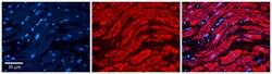
- Immunohistochemical analysis of PKA alpha in paraffin-embedded human adult heart tissue. Sample was probed with a PKA alpha polyclonal antibody (Product # PA5-70360) at a dilution of 1:600. Detection was performed with a donkey anti-Rabbit Cy3 secondary antibody at a dilution of 1:200. Images were taken at 20x magnification with an exposure time of 0.5-2.0 seconds.

- Submitted by
- Invitrogen Antibodies (provider)
- Main image
- Experimental details
- Immunohistochemical analysis of PKA alpha in paraffin-embedded human adult heart tissue. Sample was probed with a PKA alpha polyclonal antibody (Product # PA5-70360) at a dilution of 1:600. Detection was performed with a donkey anti-Rabbit Cy3 secondary antibody at a dilution of 1:200. Images were taken at 20x magnification with an exposure time of 0.5-2.0 seconds.

Supportive validation
- Submitted by
- Invitrogen Antibodies (provider)
- Main image
- Experimental details
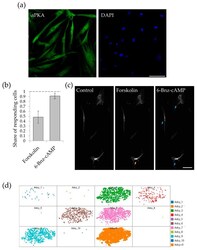
- Figure 1 Multipotent stromal cells (MSC) response to adenylate cyclase and protein kinase A (PKA) activation. ( a ) Immunofluorescent staining of the catalytic subunit of protein kinase A in MSC. Scale bar 100 um. ( b ) Share of the cells responding to adenylate cyclase activator forskolin (10 -6 M) and cell-permeable direct activator of protein kinase A 6-Bnz-cyclic AMP (10 -4 M). Results are presented as mean +- SE, n = 17-24 in 4 independent experiments on the material of 3 different donors. ( c ) Representative field of view of the cells responding to the sequential adding of forskolin and 6-Bnz-cAMP. Yellow arrow marks the cell responded to the forskolin added. Blue arrows show the cells responded to 6-Bnz-cAMP added. Scale bar 100 um. ( d ) Expression of mRNA of different isoforms of adenylyl cyclase in human MSC population. Raw data of scRNAseq analysis of MSC were downloaded from Reference [].
