 Explore
 Validate
 Learn
Learn Western blot
Antibody data
- Antibody Data
- Antigen structure
- References [1]
- Comments [0]
- Validations
- Western blot [2]
Submit
Validation data
Reference
Comment
Report error
- Product number
- MAB5908 - Provider product page

- Provider
- R&D Systems
- Product name
- Human/Mouse/Rat PKA C alpha/beta Antibody
- Antibody type
- Monoclonal
- Description
- Protein A or G purified from hybridoma culture supernatant. Detects human, mouse, and rat PKA C alpha/beta in Western blots.
- Reactivity
- Human, Mouse, Rat
- Host
- Mouse
- Conjugate
- Unconjugated
- Antigen sequence
P17612- Isotype
- IgG
- Antibody clone number
- 515741
- Vial size
- 100 ug
- Concentration
- LYOPH
- Storage
- Use a manual defrost freezer and avoid repeated freeze-thaw cycles. 12 months from date of receipt, -20 to -70 °C as supplied. 1 month, 2 to 8 °C under sterile conditions after reconstitution. 6 months, -20 to -70 °C under sterile conditions after reconstitution.
Submitted references cAMP initiates early phase neuron-like morphology changes and late phase neural differentiation in mesenchymal stem cells.
Zhang L, Seitz LC, Abramczyk AM, Liu L, Chan C
Cellular and molecular life sciences : CMLS 2011 Mar;68(5):863-76
Cellular and molecular life sciences : CMLS 2011 Mar;68(5):863-76
No comments: Submit comment
Supportive validation
- Submitted by
- R&D Systems (provider)
- Main image
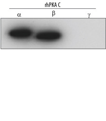
- Experimental details
- Detection of Human PKA C alpha/beta by Western Blot. Western blot shows Recombinant Human Active PKA C alpha (Catalog # 4268-KS), Recombinant Human Active PKA C beta (Catalog # 4596-KS), and recombinant human PKA C gamma (1 ng/lane each). PVDF membrane was probed with 0.2 µg/mL Mouse Anti-Human/Mouse/Rat PKA C alpha/beta Monoclonal Antibody (Catalog # MAB5908) followed by HRP-conjugated Anti-Mouse IgG Secondary Antibody (Catalog # HAF007). This experiment was conducted under reducing conditions and using Immunoblot Buffer Group 3.

- Submitted by
- R&D Systems (provider)
- Main image
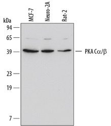
- Experimental details
- Detection of Human/Mouse/Rat PKA C alpha/beta by Western Blot. Western blot shows lysates of MCF-7 human breast cancer cell line, Neuro-2A mouse neuroblastoma cell line, and Rat-2 rat embryonic fibroblast cell line. PVDF membrane was probed with 0.2 µg/mL Mouse Anti-Human/Mouse/Rat PKA C alpha/beta Monoclonal Antibody (Catalog # MAB5908) followed by HRP-conjugated Anti-Mouse IgG Secondary Antibody (Catalog # HAF007). A specific band for PKA C alpha/beta was detected at approximately 40 kDa (as indicated). This experiment was conducted under reducing conditions and using Immunoblot Buffer Group 3.
