 Explore
 Validate
 Learn
Learn Western blot
Antibody data
- Antibody Data
- Antigen structure
- References [0]
- Comments [0]
- Validations
- Western blot [2]
- Immunohistochemistry [1]
Submit
Validation data
Reference
Comment
Report error
- Product number
- PA5-48072 - Provider product page

- Provider
- Invitrogen Antibodies
- Product name
- DACH2 Polyclonal Antibody
- Antibody type
- Polyclonal
- Antigen
- Recombinant full-length protein
- Description
- This antibody detects endogenous human and mouse DACH2 in Western blots.
- Concentration
- 0.2 mg/mL
No comments: Submit comment
Supportive validation
- Submitted by
- Invitrogen Antibodies (provider)
- Main image

- Experimental details
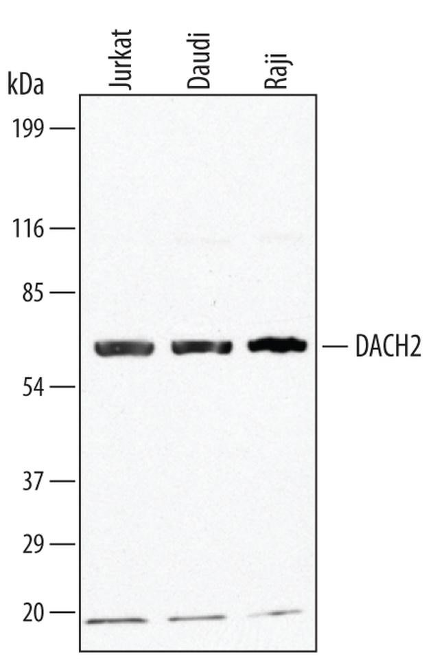
- Western blot analysis from lysates of Jurkat human acute T cell leukemia cell line, Daudi human Burkitt's lymphoma cell line, and Raji human Burkitt's lymphoma cell line. PVDF membrane was probed with 1 µg/mL of human/mouse DACH2 Antigen Affinity-purified Polyclonal Antibody (Product # PA5-48072) followed by HRP-conjugated Anti-Goat IgG Secondary Antibody. A specific band was detected for DACH2 at approximately 65 kDa (as indicated). This experiment was conducted under reducing conditions.
- Submitted by
- Invitrogen Antibodies (provider)
- Main image

- Experimental details
- Western blot analysis of DACH2 in Jurkat human acute T cell leukemia cell line, Daudi human Burkitts lymphoma cell line, and Raji human Burkitts lymphoma cell line. Samples were incubated in DACH2 polyclonal antibody (Product # PA5-48072) using a dilution of 1 µg/mL followed by a HRP-conjugated Anti-Goat IgG secondary antibody. A specific band was detected for DACH2 at approximately 65 kDa (as indicated). This experiment was conducted under reducing conditions.

Supportive validation
- Submitted by
- Invitrogen Antibodies (provider)
- Main image

- Experimental details
- Immunohistochemical analysis of DACH2 in perfusion fixed frozen sections of mouse embryonic brain (13 d.p.c.). Samples were incubated with DACH2 polyclonal antibody (Product # PA5-48072) using a dilution of 5 µg/mL for 1 hour at room temperature followed by Anti-Goat IgG VisUCyte™ HRP Polymer Antibody. Tissue was stained using DAB (brown) and counterstained with hematoxylin (blue). Specific staining was localized to nuclei in neuronal cells.
