 Explore
 Validate
 Learn
Learn Western blot
Antibody data
- Antibody Data
- Antigen structure
- References [0]
- Comments [0]
- Validations
- Western blot [6]
- Immunocytochemistry [1]
- Immunohistochemistry [1]
Submit
Validation data
Reference
Comment
Report error
- Product number
- PA5-27752 - Provider product page

- Provider
- Invitrogen Antibodies
- Product name
- DDR2 Polyclonal Antibody
- Antibody type
- Polyclonal
- Antigen
- Recombinant protein fragment
- Description
- Recommended positive controls: Raji, mouse brain.
- Concentration
- 0.43 mg/mL
No comments: Submit comment
Supportive validation
- Submitted by
- Invitrogen Antibodies (provider)
- Main image

- Experimental details
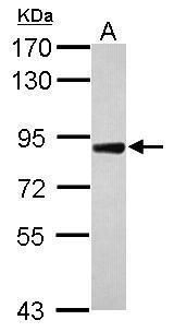
- Western blot analysis of DDR2 using 30 µg of Raji lysate. Samples were loaded onto a 7.5% SDS-PAGE gel and probed with a DDR2 polyclonal antibody (Product # PA5-27752) at a dilution of 1:5000.
- Submitted by
- Invitrogen Antibodies (provider)
- Main image
- Experimental details
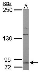
- Western blot analysis of DDR2 using 50 µg of mouse brain lysate. Samples were loaded onto a 5% SDS-PAGE gel and probed with a DDR2 polyclonal antibody (Product # PA5-27752) at a dilution of 1:5000.

- Submitted by
- Invitrogen Antibodies (provider)
- Main image
- Experimental details
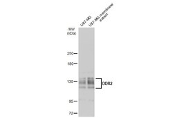
- Western Blot analysis of DDR2 was performed by separating 30 µg of U87-MG whole cell and membrane extracts by 5% SDS-PAGE. Proteins were transferred to a membrane and probed with a DDR2 Polyclonal Antibody (Product # PA5-27752) at a dilution of 1:500 and a HRP-conjugated anti-rabbit IgG secondary antibody.

- Submitted by
- Invitrogen Antibodies (provider)
- Main image
- Experimental details
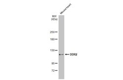
- Western Blot using DDR2 Polyclonal Antibody (Product # PA5-27752). Mouse tissue extract (50 µg) was separated by 5% SDS-PAGE, and the membrane was blotted with DDR2 Polyclonal Antibody (Product # PA5-27752) diluted at 1:1,000. The HRP-conjugated anti-rabbit IgG antibody was used to detect the primary antibody.

- Submitted by
- Invitrogen Antibodies (provider)
- Main image
- Experimental details
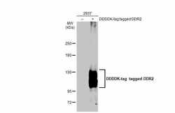
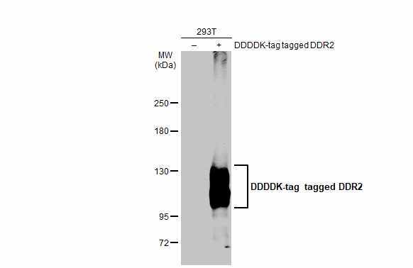
- Western Blot using DDR2 Polyclonal Antibody (Product # PA5-27752). Non-transfected (–) and transfected (+) 293T whole cell extracts (30 µg) were separated by 5% SDS-PAGE, and the membrane was blotted with DDR2 Polyclonal Antibody (Product # PA5-27752) diluted at 1:3,000. The HRP-conjugated anti-rabbit IgG antibody was used to detect the primary antibody.
- Submitted by
- Invitrogen Antibodies (provider)
- Main image
- Experimental details
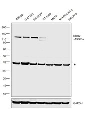
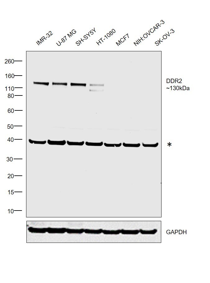
- Western blot was performed using Anti-DDR2 Polyclonal Antibody (Product # PA5-27752) and a 130kDa band corresponding to DDR2 was observed in IMR-32, U-87 MG, SH-SY5Y and HT-1080. No corresponding band of interest was observed in MCF7, NIH:OVCAR-3 and SKOV-3 which are known to be low expressing cell lines (DOI: 10.18632/oncotarget.8795). Whole cell extracts (30 µg lysate) of IMR-32 (Lane 1), U-87 MG (Lane 2), SH-SY5Y (Lane 3), HT-1080 (Lane 4), MCF7 (Lane 5), NIH:OVCAR-3 (Lane 6) and SK-O-V3 (Lane 7) were electrophoresed using NuPAGE™ 4-12% Bis-Tris Protein Gel (Product # NP0321BOX). Resolved proteins were then transferred onto a Nitrocellulose membrane (Product # IB23001) by iBlot® 2 Dry Blotting System (Product # IB21001). The blot was probed with the primary antibody (1:5000 dilution) and detected by chemiluminescence with Goat anti-Rabbit IgG (H+L) Superclonal™ Recombinant Secondary Antibody, HRP (Product # A27036, 1:4000 dilution) using the iBright FL 1000 (Product # A32752). Chemiluminescent detection was performed using Novex® ECL Chemiluminescent Substrate Reagent Kit (Product # WP20005). An uncharacterized band (*) was also observed at ~40kDa.
Supportive validation
- Submitted by
- Invitrogen Antibodies (provider)
- Main image
- Experimental details
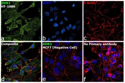
- Immunofluorescence analysis of DDR2 was performed using 70% confluent log phase HT-1080 cells. The cells were fixed with 4% paraformaldehyde for 10 minutes, permeabilized with 0.1% Triton™ X-100 for 15 minutes, and blocked with 2% BSA for 45 minutes at room temperature. The cells were labeled with DDR2 Polyclonal Antibody (Product # PA5-27752) at 1:200 dilution in 0.1% BSA, incubated at 4 degree celsius overnight and then labeled with Donkey anti-Rabbit IgG (H+L) Highly Cross-Adsorbed Secondary Antibody, Alexa Fluor Plus 488 (Product # A32790), (1:2000 dilution), for 45 minutes at room temperature (Panel a: Green). Nuclei (Panel b: Blue) were stained with ProLong™ Diamond Antifade Mountant with DAPI (Product # P36962). F-actin (Panel c: Red) was stained with Rhodamine Phalloidin (Product # R415, 1:300). Panel d represents the merged image showing plasma membrane localization. Panel e represents MCF7 cells with lower expression of DDR2 (DOI: 10.18632/oncotarget.8795). Panel f represents control cells with no primary antibody to assess background. The images were captured at 60X magnification.

Supportive validation
- Submitted by
- Invitrogen Antibodies (provider)
- Main image
- Experimental details
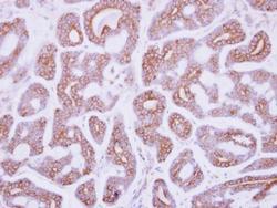
- Immunohistochemical analysis of paraffin-embedded human breast cancer, using DDR2 (Product # PA5-27752) antibody at 1:250 dilution. Antigen Retrieval: EDTA based buffer, pH 8.0, 15 min.
