Antibody data
- Antibody Data
- Antigen structure
- References [2]
- Comments [0]
- Validations
- Western blot [6]
- Immunohistochemistry [1]
- Other assay [1]
Submit
Validation data
Reference
Comment
Report error
- Product number
- PA5-77459 - Provider product page

- Provider
- Invitrogen Antibodies
- Product name
- GLUT2 Polyclonal Antibody
- Antibody type
- Polyclonal
- Antigen
- Synthetic peptide
- Description
- For reconstitution, we recommend adding 100 µL distilled water to a final antibody concentration of about 1 mg/mL. To use this carrier-free antibody for conjugation experiments, we strongly recommend performing another round of desalting. (Zeba Spin Desalting Columns, 7KMWCO, 0.5 mL, Product # 89882)
- Reactivity
- Human, Mouse, Rat
- Host
- Rabbit
- Isotype
- IgG
- Vial size
- 50 µL
- Concentration
- 0.8 mg/mL
- Storage
- -20°C
Submitted references The Postnatal Offspring of Finasteride-Treated Male Rats Shows Hyperglycaemia, Elevated Hepatic Glycogen Storage and Altered GLUT2, IR, and AR Expression in the Liver.
The Fast Lane of Hypoxic Adaptation: Glucose Transport Is Modulated via A HIF-Hydroxylase-AMPK-Axis in Jejunum Epithelium.
Kur P, Kolasa-Wołosiuk A, Grabowska M, Kram A, Tarnowski M, Baranowska-Bosiacka I, Rzeszotek S, Piasecka M, Wiszniewska B
International journal of molecular sciences 2021 Jan 27;22(3)
International journal of molecular sciences 2021 Jan 27;22(3)
The Fast Lane of Hypoxic Adaptation: Glucose Transport Is Modulated via A HIF-Hydroxylase-AMPK-Axis in Jejunum Epithelium.
Dengler F, Gäbel G
International journal of molecular sciences 2019 Oct 9;20(20)
International journal of molecular sciences 2019 Oct 9;20(20)
No comments: Submit comment
Supportive validation
- Submitted by
- Invitrogen Antibodies (provider)
- Main image

- Experimental details
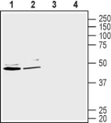
- Western blot analysis of mouse brain membranes with GLUT2 polyclonal antibody (Product # PA5-77459) using a dilution of 1:200.
- Submitted by
- Invitrogen Antibodies (provider)
- Main image

- Experimental details
- Western blot analysis of mouse brain membranes with GLUT2 polyclonal antibody (Product # PA5-77459) using a dilution of 1:200.
- Submitted by
- Invitrogen Antibodies (provider)
- Main image

- Experimental details
- Western blot analysis of mouse brain membranes with GLUT2 polyclonal antibody (Product # PA5-77459) using a dilution of 1:200.
- Submitted by
- Invitrogen Antibodies (provider)
- Main image

- Experimental details
- Western blot analysis of mouse brain membranes with GLUT2 polyclonal antibody (Product # PA5-77459) using a dilution of 1:200.
- Submitted by
- Invitrogen Antibodies (provider)
- Main image

- Experimental details
- Western blot analysis of mouse brain membranes with GLUT2 polyclonal antibody (Product # PA5-77459) using a dilution of 1:200.
- Submitted by
- Invitrogen Antibodies (provider)
- Main image

- Experimental details
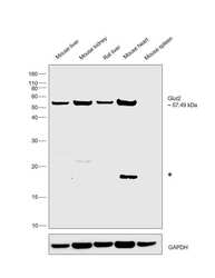
- Western blot was performed using anti-Glut2 Polyclonal Antibody (Product # PA5-77459) on tissue extracts (30 µg lysate) of Mouse liver (Lane1), Mouse kidney (Lane 2), Rat liver (Lane 3), Mouse heart (Lane 4) and Mouse spleen (Lane 5) and 57.49 kDa band corresponding to GLUT2 protein was observed. Resolved proteins were then transferred onto a nitrocellulose membrane (Product # IB23001) by iBlot® 2 Dry Blotting System (Product # IB21001). The blot was probed with the primary antibody (1:1000 dilution) and detected by chemiluminescence with Goat anti-Rabbit IgG (H+L) Superclonal™ Recombinant Secondary Antibody, HRP (Product # A27036, 0.25 µg/mL, 1:4000 dilution) using the iBright FL 1000 (Product # A32752). Chemiluminescent detection was performed using Novex® ECL Chemiluminescent Substrate Reagent Kit (Product # WP20005). (An uncharacterized band (*) was observed in few tissues tested).
Supportive validation
- Submitted by
- Invitrogen Antibodies (provider)
- Main image

- Experimental details
- Immunohistochemistry analysis of GLUT2 in perfusion-fixed, frozen rat brain. A) Samples were probed with GLUT2 polyclonal antibody (Product # PA5-77459) using a dilution of 1:120, and incubated with goat-anti-rabbit-AlexaFluor-488 (green). B) Staining in hippocampus dentate gyrus, shows labeling of both granule cells (horizontal arrow) and in hilar interneurons (vertical arrows).
Supportive validation
- Submitted by
- Invitrogen Antibodies (provider)
- Main image

- Experimental details
- Figure 7 ( A ) The glucose transporter 2 mRNA levels (normalized to GAPDH) in the homogenates of hepatic tissue of control offspring (F1:Control) and those (F1:Fin) born from females fertilized by finasteride-treated male rats in postnatal days 7, 14, 21, 28, and 90. Values are expressed as arithmetic means +- SD; differences were evaluated using the Mann-Whitney U -test ( n = 5 per each age group; p
 Explore
 Validate
 Learn
Learn Western blot