Antibody data
- Antibody Data
- Antigen structure
- References [6]
- Comments [0]
- Validations
- Western blot [1]
- Immunocytochemistry [1]
- Immunohistochemistry [6]
Submit
Validation data
Reference
Comment
Report error
- Product number
- HPA000660 - Provider product page

- Provider
- Atlas Antibodies
- Proper citation
- Atlas Antibodies Cat#HPA000660, RRID:AB_1080579
- Product name
- Anti-VRK1
- Antibody type
- Polyclonal
- Reactivity
- Human
- Host
- Rabbit
- Conjugate
- Unconjugated
- Antigen sequence
KAAQAGRQSSAKRHLAEQFAVGEIITDMAKKEWKV
GLPIGQGGFGCIYLADMNSSESVGSDAPCVVKVEP
SDNGPLFTELKFYQRAAKPEQIQKWIRTRKLKYLG
VPKYWGSGLHDKNGKSYRFMIMDRFGSDLQKI- Isotype
- IgG
- Vial size
- 100 µl
- Storage
- Store at +4°C for short term storage. Long time storage is recommended at -20°C.
Submitted references VRK1 regulates Cajal body dynamics and protects coilin from proteasomal degradation in cell cycle.
Depletion of the protein kinase VRK1 disrupts nuclear envelope morphology and leads to BAF retention on mitotic chromosomes.
Vaccinia-related kinase 1 (VRK1) confers resistance to DNA-damaging agents in human breast cancer by affecting DNA damage response.
Rewiring of human lung cell lineage and mitotic networks in lung adenocarcinomas.
Molecular genetic analysis of VRK1 in mammary epithelial cells: depletion slows proliferation in vitro and tumor growth and metastasis in vivo.
Vaccinia-related kinase 1 (VRK1) is an upstream nucleosomal kinase required for the assembly of 53BP1 foci in response to ionizing radiation-induced DNA damage.
Cantarero L, Sanz-García M, Vinograd-Byk H, Renbaum P, Levy-Lahad E, Lazo PA
Scientific reports 2015 Jun 12;5:10543
Scientific reports 2015 Jun 12;5:10543
Depletion of the protein kinase VRK1 disrupts nuclear envelope morphology and leads to BAF retention on mitotic chromosomes.
Molitor TP, Traktman P
Molecular biology of the cell 2014 Mar;25(6):891-903
Molecular biology of the cell 2014 Mar;25(6):891-903
Vaccinia-related kinase 1 (VRK1) confers resistance to DNA-damaging agents in human breast cancer by affecting DNA damage response.
Salzano M, Vázquez-Cedeira M, Sanz-García M, Valbuena A, Blanco S, Fernández IF, Lazo PA
Oncotarget 2014 Apr 15;5(7):1770-8
Oncotarget 2014 Apr 15;5(7):1770-8
Rewiring of human lung cell lineage and mitotic networks in lung adenocarcinomas.
Kim IJ, Quigley D, To MD, Pham P, Lin K, Jo B, Jen KY, Raz D, Kim J, Mao JH, Jablons D, Balmain A
Nature communications 2013;4:1701
Nature communications 2013;4:1701
Molecular genetic analysis of VRK1 in mammary epithelial cells: depletion slows proliferation in vitro and tumor growth and metastasis in vivo.
Molitor TP, Traktman P
Oncogenesis 2013 Jun 3;2(6):e48
Oncogenesis 2013 Jun 3;2(6):e48
Vaccinia-related kinase 1 (VRK1) is an upstream nucleosomal kinase required for the assembly of 53BP1 foci in response to ionizing radiation-induced DNA damage.
Sanz-García M, Monsalve DM, Sevilla A, Lazo PA
The Journal of biological chemistry 2012 Jul 6;287(28):23757-68
The Journal of biological chemistry 2012 Jul 6;287(28):23757-68
No comments: Submit comment
Enhanced validation
- Submitted by
- Atlas Antibodies (provider)
- Enhanced method
- Genetic validation
- Main image

- Experimental details
- Western blot analysis in Rh30 cells transfected with control siRNA, target specific siRNA probe #1, using Anti-VRK1 antibody. Remaining relative intensity is presented. Loading control: Anti-PPIB.
Supportive validation
- Submitted by
- Atlas Antibodies (provider)
- Main image

- Experimental details
- Immunofluorescent staining of human cell line U-251 MG shows localization to nucleoplasm.
- Sample type
- HUMAN
Enhanced validation
Supportive validation
- Submitted by
- Atlas Antibodies (provider)
- Enhanced method
- Orthogonal validation
- Main image

- Experimental details
- Immunohistochemistry analysis in human testis and pancreas tissues using HPA000660 antibody. Corresponding VRK1 RNA-seq data are presented for the same tissues.
- Sample type
- HUMAN
Supportive validation
- Submitted by
- Atlas Antibodies (provider)
- Main image

- Experimental details
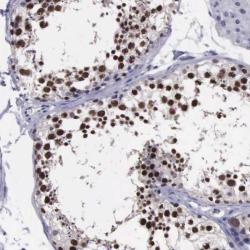
- Immunohistochemical staining of human testis shows strong nuclear positivity in cells in seminiferus ducts.
- Submitted by
- Atlas Antibodies (provider)
- Main image

- Experimental details
- Immunohistochemical staining of human testis shows strong nuclear positivity in cells in seminiferous ducts.
- Sample type
- HUMAN
- Submitted by
- Atlas Antibodies (provider)
- Main image

- Experimental details
- Immunohistochemical staining of human tonsil shows moderate nuclear positivity in lymphocytes.
- Sample type
- HUMAN
- Submitted by
- Atlas Antibodies (provider)
- Main image

- Experimental details
- Immunohistochemical staining of human placenta shows strong nuclear positivity in trophoblastic cells.
- Sample type
- HUMAN
- Submitted by
- Atlas Antibodies (provider)
- Main image

- Experimental details
- Immunohistochemical staining of human pancreas shows no positivity as expected.
- Sample type
- HUMAN
 Explore
 Validate
 Learn
Learn Western blot
 Immunocytochemistry