 Explore
 Validate
 Learn
Learn Western blot
Antibody data
- Antibody Data
- Antigen structure
- References [9]
- Comments [0]
- Validations
- Western blot [6]
- Immunocytochemistry [3]
- Immunohistochemistry [1]
Submit
Validation data
Reference
Comment
Report error
- Product number
- GTX101416 - Provider product page

- Provider
- GeneTex
- Proper citation
- GeneTex Cat#GTX101416, RRID:AB_10726413
- Product name
- LDHA antibody
- Antibody type
- Polyclonal
- Reactivity
- Human, Mouse, Rat
- Host
- Rabbit
Submitted references Proteomic analysis of evodiamine-induced cytotoxicity in thyroid cancer cells.
Transketolase Regulates the Metabolic Switch to Control Breast Cancer Cell Metastasis via the α-Ketoglutarate Signaling Pathway.
Targeting the Mevalonate Pathway Suppresses VHL-Deficient CC-RCC through an HIF-Dependent Mechanism.
Proteomic analysis of honokiol-induced cytotoxicity in thyroid cancer cells.
Thiamine deficiency activates hypoxia inducible factor-1α to facilitate pro-apoptotic responses in mouse primary astrocytes.
Interleukin-17 limits hypoxia-inducible factor 1α and development of hypoxic granulomas during tuberculosis.
Diet-induced obesity accelerates blood lactate accumulation of rats in response to incremental exercise to maximum.
Continued 26S proteasome dysfunction in mouse brain cortical neurons impairs autophagy and the Keap1-Nrf2 oxidative defence pathway.
Rho-associated kinase 1 inhibition is synthetically lethal with von Hippel-Lindau deficiency in clear cell renal cell carcinoma.
Yu HI, Chou HC, Su YC, Lin LH, Lu CH, Chuang HH, Tsai YT, Liao EC, Wei YS, Yang YT, Lee YR, Chan HL
Journal of pharmaceutical and biomedical analysis 2018 Oct 25;160:344-350
Journal of pharmaceutical and biomedical analysis 2018 Oct 25;160:344-350
Transketolase Regulates the Metabolic Switch to Control Breast Cancer Cell Metastasis via the α-Ketoglutarate Signaling Pathway.
Tseng CW, Kuo WH, Chan SH, Chan HL, Chang KJ, Wang LH
Cancer research 2018 Jun 1;78(11):2799-2812
Cancer research 2018 Jun 1;78(11):2799-2812
Targeting the Mevalonate Pathway Suppresses VHL-Deficient CC-RCC through an HIF-Dependent Mechanism.
Thompson JM, Alvarez A, Singha MK, Pavesic MW, Nguyen QH, Nelson LJ, Fruman DA, Razorenova OV
Molecular cancer therapeutics 2018 Aug;17(8):1781-1792
Molecular cancer therapeutics 2018 Aug;17(8):1781-1792
Proteomic analysis of honokiol-induced cytotoxicity in thyroid cancer cells.
Chou HC, Lu CH, Su YC, Lin LH, Yu HI, Chuang HH, Tsai YT, Liao EC, Wei YS, Yang YT, Chien YA, Yu XR, Lee YR, Chan HL
Life sciences 2018 Aug 15;207:184-204
Life sciences 2018 Aug 15;207:184-204
Thiamine deficiency activates hypoxia inducible factor-1α to facilitate pro-apoptotic responses in mouse primary astrocytes.
Zera K, Zastre J
PloS one 2017;12(10):e0186707
PloS one 2017;12(10):e0186707
Interleukin-17 limits hypoxia-inducible factor 1α and development of hypoxic granulomas during tuberculosis.
Domingo-Gonzalez R, Das S, Griffiths KL, Ahmed M, Bambouskova M, Gopal R, Gondi S, Muñoz-Torrico M, Salazar-Lezama MA, Cruz-Lagunas A, Jiménez-Álvarez L, Ramirez-Martinez G, Espinosa-Soto R, Sultana T, Lyons-Weiler J, Reinhart TA, Arcos J, de la Luz Garcia-Hernandez M, Mastrangelo MA, Al-Hammadi N, Townsend R, Balada-Llasat JM, Torrelles JB, Kaplan G, Horne W, Kolls JK, Artyomov MN, Rangel-Moreno J, Zúñiga J, Khader SA
JCI insight 2017 Oct 5;2(19)
JCI insight 2017 Oct 5;2(19)
Diet-induced obesity accelerates blood lactate accumulation of rats in response to incremental exercise to maximum.
Chen CJ, Liao YH, Lin SY, Yu JX, Li ZJ, Lin YC, Chang GJ, Lin CH, Wong AM
American journal of physiology. Regulatory, integrative and comparative physiology 2017 Nov 1;313(5):R601-R607
American journal of physiology. Regulatory, integrative and comparative physiology 2017 Nov 1;313(5):R601-R607
Continued 26S proteasome dysfunction in mouse brain cortical neurons impairs autophagy and the Keap1-Nrf2 oxidative defence pathway.
Ugun-Klusek A, Tatham MH, Elkharaz J, Constantin-Teodosiu D, Lawler K, Mohamed H, Paine SM, Anderson G, John Mayer R, Lowe J, Ellen Billett E, Bedford L
Cell death & disease 2017 Jan 5;8(1):e2531
Cell death & disease 2017 Jan 5;8(1):e2531
Rho-associated kinase 1 inhibition is synthetically lethal with von Hippel-Lindau deficiency in clear cell renal cell carcinoma.
Thompson JM, Nguyen QH, Singh M, Pavesic MW, Nesterenko I, Nelson LJ, Liao AC, Razorenova OV
Oncogene 2017 Feb 23;36(8):1080-1089
Oncogene 2017 Feb 23;36(8):1080-1089
No comments: Submit comment
Supportive validation
- Submitted by
- GeneTex (provider)
- Main image

- Experimental details
- Sample (30 ug of whole cell lysate) A: A431 10% SDS PAGE GTX101416 diluted at 1:1000
- Validation comment
- WB

- Submitted by
- GeneTex (provider)
- Main image

- Experimental details
- LDHA antibody detects LDHA protein by western blot analysis.A. 30 ?g PC-12 whole cell lysate/extractB. 30 ?g Rat2 whole cell lysate/extract10% SDS-PAGELDHA antibody (GTX101416) dilution: 1:1000 The HRP-conjugated anti-rabbit IgG antibody (GTX213110-01) was used to detect the primary antibody.

- Submitted by
- GeneTex (provider)
- Main image

- Experimental details
- LDHA antibody detects LDHA protein by western blot analysis.A. 30 ?g Neuro2A whole cell lysate/extract B. 30 ?g C8D30 whole cell lysate/extract C. 30 ?g NIH-3T3 whole cell lysate/extract D. 30 ?g Raw264.7 whole cell lysate/extract E. 30 ?g C2C12 whole cell lysate/extract10% SDS-PAGELDHA antibody (GTX101416) dilution: 1:1000 The HRP-conjugated anti-rabbit IgG antibody (GTX213110-01) was used to detect the primary antibody.

- Submitted by
- GeneTex (provider)
- Main image
- Experimental details
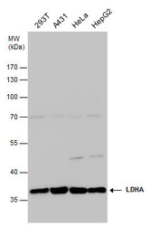
- LDHA antibody detects LDHA protein by western blot analysis. Various whole cell extracts (30 ?g) were separated by 10% SDS-PAGE, and the membrane was blotted with LDHA antibody (GTX101416) diluted at 1:1000. The HRP-conjugated anti-rabbit IgG antibody (GTX213110-01) was used to detect the primary antibody.

- Submitted by
- GeneTex (provider)
- Main image

- Experimental details
- Various whole cell extracts (30 ?g) were separated by 10% SDS-PAGE, and the membrane was blotted with LDHA antibody (GTX101416) diluted at 1:1000. The HRP-conjugated anti-rabbit IgG antibody (GTX213110-01) was used to detect the primary antibody.

- Submitted by
- GeneTex (provider)
- Main image

- Experimental details
- Various whole cell extracts (30 ?g) were separated by 12% SDS-PAGE, and the membrane was blotted with LDHA antibody (GTX101416) diluted at 1:1000.

Supportive validation
- Submitted by
- GeneTex (provider)
- Main image
- Experimental details
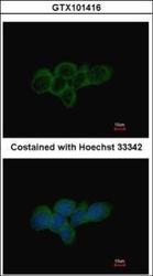
- Immunofluorescence analysis of methanol-fixed A431, using LDHA(GTX101416) antibody at 1:500 dilution.

- Submitted by
- GeneTex (provider)
- Main image

- Experimental details
- LDHA antibody detects LDHA protein at cytoplasm by immunofluorescent analysis.Sample: MCF7 cells were fixed in 4% paraformaldehyde at RT for 15 min.Green: LDHA protein stained by LDHA antibody (GTX101416) diluted at 1:1000.Red: phalloidin, a cytoskeleton marker, stained by phalloidin (invitrogen, A12380) diluted at 1:200.Blue: Hoechst 33342 staining.

- Submitted by
- GeneTex (provider)
- Main image

- Experimental details
- LDHA antibody detects LDHA protein at cytoplasm by immunofluorescent analysis.Sample: HeLa cells were fixed in 4% paraformaldehyde at RT for 15 min.Green: LDHA protein stained by LDHA antibody (GTX101416) diluted at 1:1000.Red: phalloidin, a cytoskeleton marker, stained by phalloidin (invitrogen, A12380) diluted at 1:200.Blue: Hoechst 33342 staining.

Supportive validation
- Submitted by
- GeneTex (provider)
- Main image

- Experimental details
- Immunohistochemical analysis of paraffin-embedded human lung papillory adenocarcinoma, using LDHA(GTX101416) antibody at 1:500 dilution.
