Antibody data
- Antibody Data
- Antigen structure
- References [2]
- Comments [0]
- Validations
- Western blot [3]
- Immunocytochemistry [3]
- Immunohistochemistry [3]
- Other assay [1]
Submit
Validation data
Reference
Comment
Report error
- Product number
- PA5-95727 - Provider product page

- Provider
- Invitrogen Antibodies
- Product name
- GM130 Polyclonal Antibody
- Antibody type
- Polyclonal
- Antigen
- Recombinant full-length protein
- Description
- Positive Samples: HepG2, K562, mouse liver, Rat brain; Cellular Location: Cytoplasm, Golgi apparatus, Peripheral membrane protein, cis-Golgi network membrane, cytoskeleton, spindle pole
- Reactivity
- Human, Mouse, Rat
- Host
- Rabbit
- Isotype
- IgG
- Vial size
- 100 µL
- Concentration
- 2.41 mg/mL
- Storage
- -20° C, Avoid Freeze/Thaw Cycles
Submitted references The Synthetic Cannabinoid WIN55,212-2 Can Disrupt the Golgi Apparatus Independent of Cannabinoid Receptor-1.
Phosphatidylinositol 4-kinase III beta regulates cell shape, migration, and focal adhesion number.
Lott J, Jutkiewicz EM, Puthenveedu MA
Molecular pharmacology 2022 May;101(5):371-380
Molecular pharmacology 2022 May;101(5):371-380
Phosphatidylinositol 4-kinase III beta regulates cell shape, migration, and focal adhesion number.
Bilodeau P, Jacobsen D, Law-Vinh D, Lee JM
Molecular biology of the cell 2020 Aug 1;31(17):1904-1916
Molecular biology of the cell 2020 Aug 1;31(17):1904-1916
No comments: Submit comment
Supportive validation
- Submitted by
- Invitrogen Antibodies (provider)
- Main image

- Experimental details
- Western blot analysis of extracts of various cell lines, using GOLGA2 Polyclonal antibody (Product # PA5-95727) at 1:1000 dilution. Secondary antibody: HRP Goat Anti-Rabbit IgG (H+L) at 1:10000 dilution. Lysates/proteins: 25ug per lane. Blocking buffer: 3% nonfat dry milk in TBST. Exposure time: 10s.
- Submitted by
- Invitrogen Antibodies (provider)
- Main image

- Experimental details
- Western blot analysis of extracts of rat brain, using GOLGA2 Polyclonal antibody (Product # PA5-95727) at 1:1000 dilution. Secondary antibody: HRP Goat Anti-Rabbit IgG (H+L) at 1:10000 dilution. Lysates/proteins: 25ug per lane. Blocking buffer: 3% nonfat dry milk in TBST. Exposure time: 90s.
- Submitted by
- Invitrogen Antibodies (provider)
- Main image

- Experimental details
- Western Blot analysis of GM130 in extracts of various cell lines using GM130 Polyclonal Antibody (Product # PA5-95727) at a dilution of 1:1000. A HRP Goat Anti-Rabbit IgG (H+L) secondary antibody was used at a dilution of 1:10,000. Lysates/proteins: 25 µg per lane. Blocking buffer: 3% nonfat dry milk in TBST.
Supportive validation
- Submitted by
- Invitrogen Antibodies (provider)
- Main image

- Experimental details
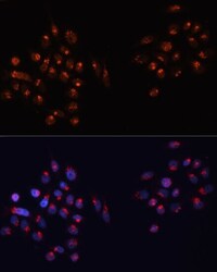
- Immunocytochemistry-Immunofluorescence analysis of GM130 was performed in HeLa cells using GM130 Polyclonal Antibody (Product # PA5-95727).
- Submitted by
- Invitrogen Antibodies (provider)
- Main image

- Experimental details
- Immunocytochemistry-Immunofluorescence analysis of GM130 was performed in HeLa cells using GM130 Polyclonal Antibody (Product # PA5-95727).
- Submitted by
- Invitrogen Antibodies (provider)
- Main image

- Experimental details
- Immunocytochemistry-Immunofluorescence analysis of GM130 was performed in U-2 OS cells using GM130 Polyclonal Antibody (Product # PA5-95727).
Supportive validation
- Submitted by
- Invitrogen Antibodies (provider)
- Main image

- Experimental details
- Immunohistochemistry analysis of GM130 in paraffin-embedded human gastric cancer using GM130 Polyclonal Antibody (Product # PA5-95727) at a dilution of 1:200.
- Submitted by
- Invitrogen Antibodies (provider)
- Main image

- Experimental details
- Immunohistochemistry analysis of GM130 in paraffin-embedded mouse lung using GM130 Polyclonal Antibody (Product # PA5-95727) at a dilution of 1:200.
- Submitted by
- Invitrogen Antibodies (provider)
- Main image

- Experimental details
- Immunohistochemistry analysis of GM130 in paraffin-embedded mouse brain using GM130 Polyclonal Antibody (Product # PA5-95727) at a dilution of 1:200.
Supportive validation
- Submitted by
- Invitrogen Antibodies (provider)
- Main image

- Experimental details
- FIGURE 4: Loss of PI4KIIIbeta alters stress fiber appearance. (A) Cis- and trans-Golgi structure as visualized by GM130 and TGN46 staining, respectively, in WT NIH3T3 cells and two independent lines of PI4KIIIbeta-deleted cells. (B) Golgi, centrosome (gamma-tubulin) and microtubule (alpha-tubulin) structure visualized in cells entering a scratch wound. (C) CRISPR cell lines both show significantly ( t test, p < 0.0001) more cells with high numbers of stress fibers compared with WT cells. Representative cells with respectively low or high numbers of stress fibers (yellow arrows) are shown in the right panels. Bottom panels show representative fields of WT and CRISPR lines showing cells with high or low stress fiber content.
 Explore
 Validate
 Learn
Learn Western blot