 Explore
 Validate
 Learn
Learn Western blot
Antibody data
- Antibody Data
- Antigen structure
- References [0]
- Comments [0]
- Validations
- Western blot [1]
- Immunocytochemistry [1]
- Immunohistochemistry [2]
- Flow cytometry [2]
Submit
Validation data
Reference
Comment
Report error
- Product number
- MA5-32694 - Provider product page

- Provider
- Invitrogen Antibodies
- Product name
- MRP1 Recombinant Rabbit Monoclonal Antibody (JA93-03)
- Antibody type
- Monoclonal
- Antigen
- Synthetic peptide
- Reactivity
- Human
- Host
- Rabbit
- Isotype
- IgG
- Antibody clone number
- JA93-03
- Vial size
- 100 µL
- Concentration
- 1 mg/mL
- Storage
- Store at 4°C short term. For long term storage, store at -20°C, avoiding freeze/thaw cycles.
No comments: Submit comment
Supportive validation
- Submitted by
- Invitrogen Antibodies (provider)
- Main image
- Experimental details
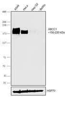
- Western blot was performed using Anti-MRP1 Recombinant Rabbit Monoclonal Antibody (JA93-03) (Product # MA5-32694) and a 150-220 kDa band corresponding to ABCC1 was observed across cell lines tested. Membrane enriched extracts (40 µg lysate) of A549 (Lane 1), HeLa (Lane 2), Hep G2 (Lane 3), BeWo (Lane 4) were electrophoresed using NuPAGE™ 3-8% Tris-Acetate Protein Gel (Product # EA0378BOX). Resolved proteins were then transferred onto a nitrocellulose membrane (Product # IB23001) by iBlot® 2 Dry Blotting System (Product # IB21001). The blot was probed with the primary antibody (1:500) and detected by chemiluminescence with Goat anti-Rabbit IgG (H+L) Superclonal™ Recombinant Secondary Antibody, HRP (Product # A27036,1:4000) using the iBright™ FL1500 Imaging System (Product # A44115). Chemiluminescentdetection was performed using Novex® ECL Chemiluminescent Substrate Reagent Kit (Product # WP20005).
Supportive validation
- Submitted by
- Invitrogen Antibodies (provider)
- Main image
- Experimental details
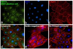
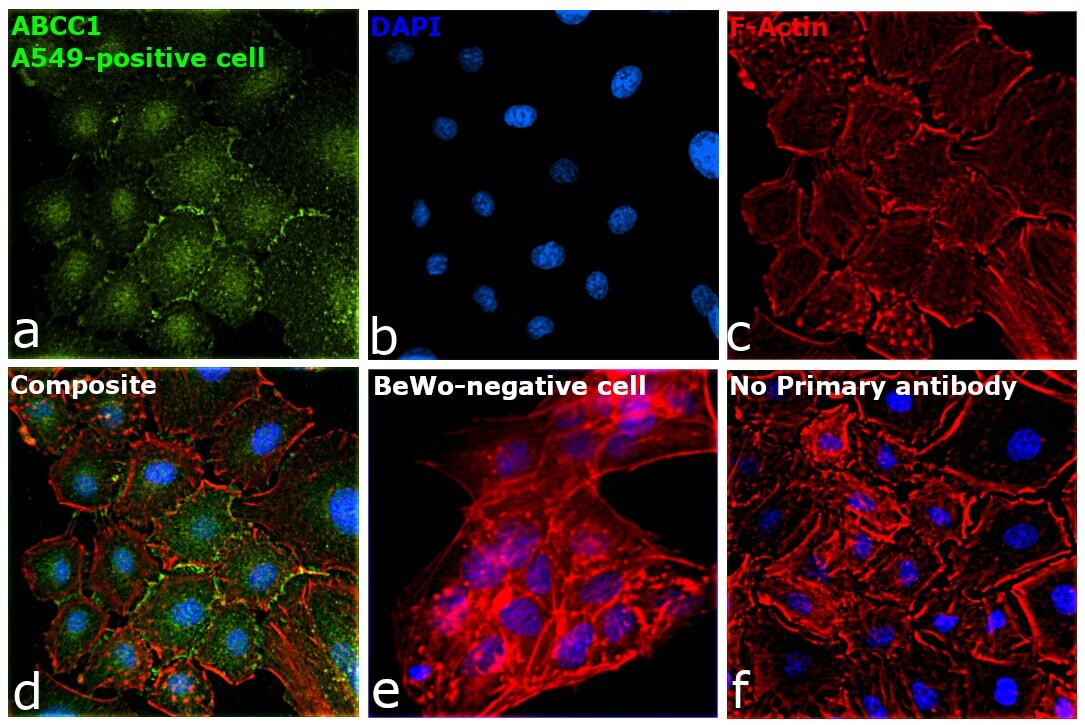
- Immunofluorescence analysis of ABCC1 was performed using 70% confluent log phase A549 cells. The cells were fixed with 4% paraformaldehyde for 10 minutes, permeabilized with 0.1% Triton™ X-100 for 10 minutes, and blocked with 2% BSA for 45 minutes at room temperature. The cells were labeled with MRP1 Recombinant Rabbit Monoclonal Antibody (JA93-03) (Product # MA5-32694) at 1:200 in 0.1% BSA, incubated at 4 degree celsius overnight and then labeled with Donkey anti-Rabbit IgG (H+L) Highly Cross-Adsorbed Secondary Antibody, Alexa Fluor Plus 488 (Product # A32790), (1:2500), for 45 minutes at room temperature (Panel a: Green). Nuclei (Panel b:Blue) were stained with ProLong™ Diamond Antifade Mountant with DAPI (Product # P36962). F-actin (Panel c: Red) was stained with Rhodamine Phalloidin (Product # R415, 1:300). Panel d represents the merged image showing plasma membrane, cell junction and nuclear localization. Panel e represents the negative expression model, BeWo. Panel f represents control cells with no primary antibody to assess background. The images were captured at 60X magnification.
Supportive validation
- Submitted by
- Invitrogen Antibodies (provider)
- Main image
- Experimental details
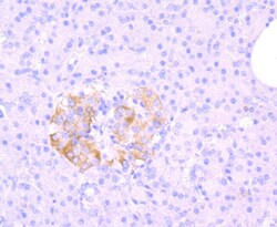
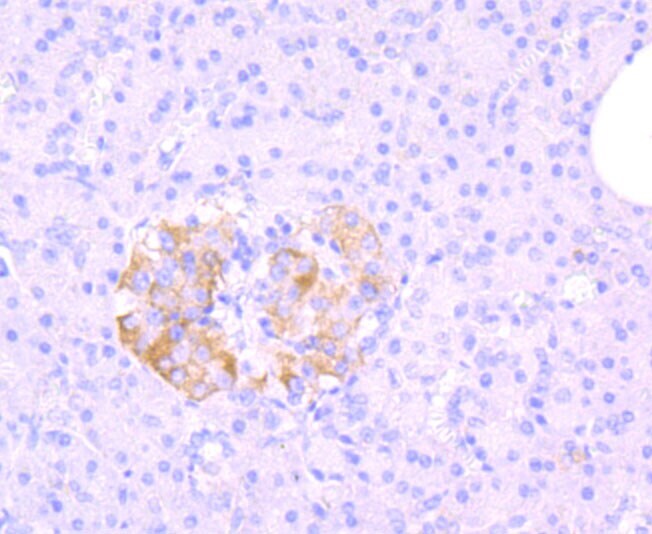
- Immunohistochemical analysis of MRP1 of paraffin-embedded Human pancreas tissue using a MRP1 Monoclonal antibody (Product #MA5-32694).
- Submitted by
- Invitrogen Antibodies (provider)
- Main image
- Experimental details
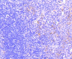
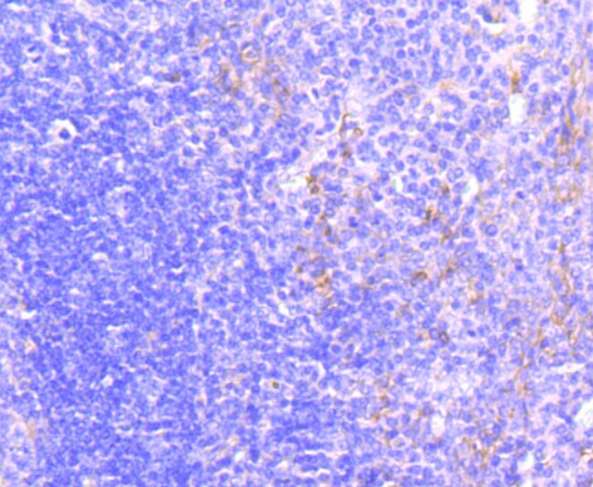
- Immunohistochemical analysis of MRP1 of paraffin-embedded Human tonsil tissue using a MRP1 Monoclonal antibody (Product #MA5-32694). Counter stained with hematoxylin.
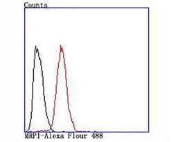
Supportive validation
- Submitted by
- Invitrogen Antibodies (provider)
- Main image
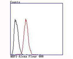
- Experimental details
- Flow Cytometric analysis of MRP1 in Hela cells using a MRP1 Monoclonal Antibody (Product # MA5-32694) at a dilution of 1:100, as seen in red compared with an unlabelled control (cells without incubation with primary antibody; black).
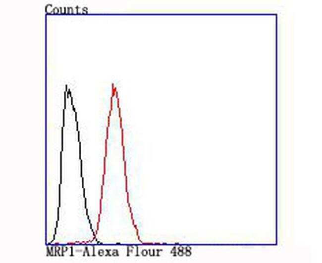
- Submitted by
- Invitrogen Antibodies (provider)
- Main image
- Experimental details
- Flow Cytometric analysis of MRP1 in Hela cells using a MRP1 Monoclonal Antibody (Product # MA5-32694) at a dilution of 1:100, as seen in red compared with an unlabelled control (cells without incubation with primary antibody; black).
