 Explore
 Validate
 Learn
Learn Western blot
 Flow cytometry
Antibody data
- Antibody Data
- Antigen structure
- References [2]
- Comments [0]
- Validations
- Western blot [1]
- Immunocytochemistry [2]
Submit
Validation data
Reference
Comment
Report error
- Product number
- MAB2506 - Provider product page

- Provider
- R&D Systems
- Product name
- Human/Mouse/Rat GSK-3 beta Antibody
- Antibody type
- Monoclonal
- Description
- Protein A or G purified from hybridoma culture supernatant. Detects human, mouse, and rat GSK-3 beta in ELISAs and Western blots. Does not cross-react with recombinant human GSK-3 alpha .
- Reactivity
- Human, Mouse, Rat
- Host
- Rat
- Conjugate
- Unconjugated
- Antigen sequence
P49841- Isotype
- IgG
- Antibody clone number
- 272536
- Vial size
- 100 ug
- Concentration
- LYOPH
- Storage
- Use a manual defrost freezer and avoid repeated freeze-thaw cycles. 12 months from date of receipt, -20 to -70 °C as supplied. 1 month, 2 to 8 °C under sterile conditions after reconstitution. 6 months, -20 to -70 °C under sterile conditions after reconstitution.
Submitted references 7,8-Dihydroxyflavone Ameliorates Cognitive Impairment by Inhibiting Expression of Tau Pathology in ApoE-Knockout Mice.
beta-catenin mediates insulin-like growth factor-I actions to promote cyclin D1 mRNA expression, cell proliferation and survival in oligodendroglial cultures.
Tan Y, Nie S, Zhu W, Liu F, Guo H, Chu J, Cao XB, Jiang X, Zhang Y, Li Y
Frontiers in aging neuroscience 2016;8:287
Frontiers in aging neuroscience 2016;8:287
beta-catenin mediates insulin-like growth factor-I actions to promote cyclin D1 mRNA expression, cell proliferation and survival in oligodendroglial cultures.
Ye P, Hu Q, Liu H, Yan Y, D'ercole AJ
Glia 2010 Jul;58(9):1031-41
Glia 2010 Jul;58(9):1031-41
No comments: Submit comment
Supportive validation
- Submitted by
- R&D Systems (provider)
- Main image
- Experimental details
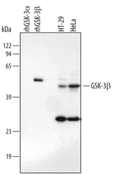
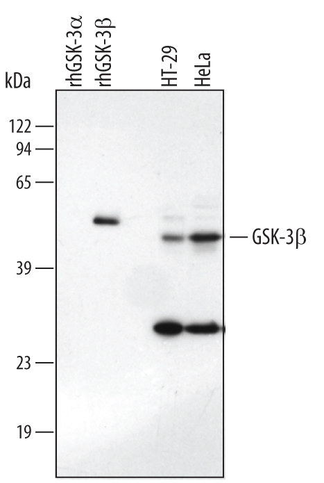
- Detection of Human/Mouse/Rat GSK-3 beta by Western Blot. Western blot shows lysates of HT-29 human colon adenocarcinoma cell line and HeLa human cervical epithelial carcinoma cell line. PVDF membrane was probed with 1 µg/mL Human/Mouse/Rat GSK-3 beta Monoclonal Antibody (Catalog # MAB2506) followed by HRP-conjugated Anti-Rat IgG Secondary Antibody (Catalog # HAF005). For additional reference, recombinant human GSK-3 alpha and Recombinant Human Active GSK-3 beta (Catalog # 2506-KS) (20 ng/lane) were included. A specific band for GSK-3 beta was detected at approximately 46 kDa (as indicated). This experiment was conducted under reducing conditions and using Immunoblot Buffer Group 1.
Supportive validation
- Submitted by
- R&D Systems (provider)
- Main image
- Experimental details
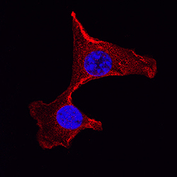
- GSK-3 beta in NIH-3T3 Mouse Cell Line. GSK-3 beta was detected in immersion fixed NIH-3T3 mouse embryonic fibroblast cell line using Rat Anti-Human/Mouse/Rat GSK-3 beta Monoclonal Antibody (Catalog # MAB2506) at 3 µg/mL for 3 hours at room temperature. Cells were stained using the NorthernLights™ 557-conjugated Anti-Rat IgG Secondary Antibody (red; Catalog # NL013) and counterstained with DAPI (blue). Specific staining was localized to nuclei and cytoplasm. View our protocol for Fluorescent ICC Staining of Cells on Coverslips.

- Submitted by
- R&D Systems (provider)
- Main image
- Experimental details
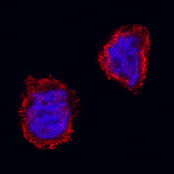
- GSK-3 beta in HT-29 Human Cell Line. GSK-3 beta was detected in immersion fixed HT-29 human colon adenocarcinoma cell line using Rat Anti-Human/Mouse/Rat GSK-3 beta Monoclonal Antibody (Catalog # MAB2506) at 3 µg/mL for 3 hours at room temperature. Cells were stained using the NorthernLights™ 557-conjugated Anti-Rat IgG Secondary Antibody (red; Catalog # NL013) and counterstained with DAPI (blue). Specific staining was localized to nuclei and cytoplasm. View our protocol for Fluorescent ICC Staining of Cells on Coverslips.
