Explore
 Validate
 Learn
Learn Western blot
Antibody data
- Antibody Data
- Antigen structure
- References [1]
- Comments [0]
- Validations
- Western blot [1]
- Immunocytochemistry [1]
- Immunohistochemistry [1]
- Other assay [2]
Submit
Validation data
Reference
Comment
Report error
- Product number
- PA5-100951 - Provider product page

- Provider
- Invitrogen Antibodies
- Product name
- CXCR2 Polyclonal Antibody
- Antibody type
- Polyclonal
- Antigen
- Synthetic peptide
- Reactivity
- Human, Mouse
- Host
- Rabbit
- Isotype
- IgG
- Vial size
- 100 µL
- Concentration
- 1 mg/mL
- Storage
- -20°C
Submitted references Loss of H2R Signaling Disrupts Neutrophil Homeostasis and Promotes Inflammation-Associated Colonic Tumorigenesis in Mice.
Shi Z, Mori-Akiyama Y, Du W, Fultz R, Zhao Y, Ruan W, Venable S, Engevik MA, Versalovic J
Cellular and molecular gastroenterology and hepatology 2022;13(3):717-737
Cellular and molecular gastroenterology and hepatology 2022;13(3):717-737
No comments: Submit comment
Supportive validation
- Submitted by
- Invitrogen Antibodies (provider)
- Main image
- Experimental details
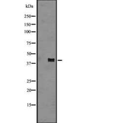
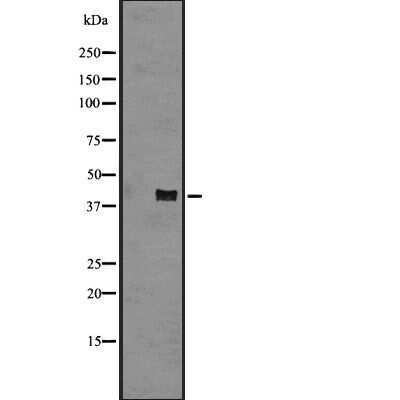
- Western blot analysis of CXCR2 in human carcinoma lysate (left lane: treated with the antigen-specific peptide). Samples were incubated with CXCR2 polyclonal antibody (Product # PA5-100951).
Supportive validation
- Submitted by
- Invitrogen Antibodies (provider)
- Main image
- Experimental details
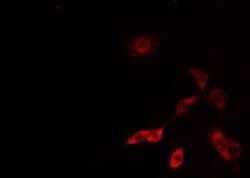
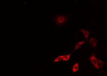
- Immunofluorescent analysis of CXCR2 in NIH-3T3. Samples were fixed with paraformaldehyde, permeabilized with 0.1% Triton X-100, blocked with 10% serum (45 min at 25°C) incubated with CXCR2 polyclonal antibody (Product # PA5-100951) using a dilution of 1:200 (1 hr, 37°C), and followed by goat anti-rabbit IgG Alexa Fluor 594 at a dilution of 1:600.
Supportive validation
- Submitted by
- Invitrogen Antibodies (provider)
- Main image
- Experimental details
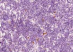
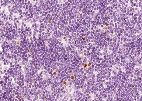
- Immunohistochemistry analysis of paraffin-embedded CXCR2 in human Lymphoma tissue sections. Antigen retrieval was performed using citrate buffer. Samples were blocked with blocking buffer (1.5 hr, 22°C), incubated with CXCR2 polyclonal antibody (Product # PA5-100951) using a dilution of 1:100 (1.5 hr, 22°C), followed by HRP conjugated goat anti-rabbit.
Supportive validation
- Submitted by
- Invitrogen Antibodies (provider)
- Main image
- Experimental details
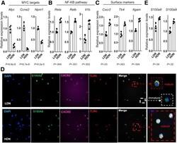
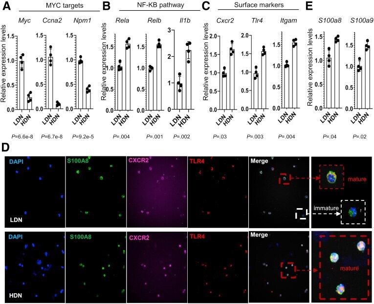
- Figure 5 Confirmation of the distinct gene signatures of LDNs and HDNs. ( A-C ) Expression of representative genes in MYC target, NF-kappaB pathways, and surface markers of LDNs and HDNs by qRT-PCR (n = 4). Means +- SD are shown. The P value is shown under each panel. ( D ) Immunofluorescence staining (magnification: 200x and 1000x) of S100A8 (green), CXCR2 (pink), and TLR4 (red) in LDNs and HDNs. ( E ) The expression levels of S100a8 and S100a9 in LDNs and HDNs by qRT-PCR (n = 4). Means +- SD are shown. P values are shown on the panel. CXCR2, C-X-C Motif Chemokine Receptor 2; DAPI, 4',6-diamidino-2-phenylindole.
- Submitted by
- Invitrogen Antibodies (provider)
- Main image
- Experimental details
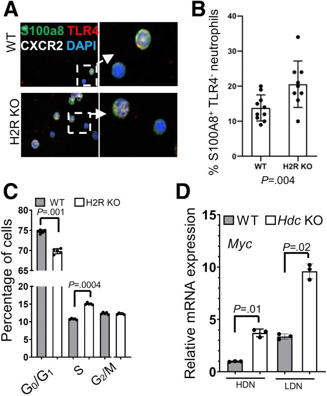
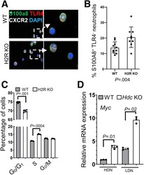
- Figure 8 H2R deficiency promoted DNA synthesis in bone marrow cells. ( A ) Immunofluorescence staining (magnification: 200x and 1000x) of S100A8 (green), CXCR2 (white), and TLR4 (red) in WT and H2R-deficient bone marrow cells. ( B ) The mean numbers in each field of TLR4-negative neutrophils at 400x power (n = 10). ( C ) DNA content of different phases of bone marrow cells determined by flow cytometry (n = 5). ( D ) The expression levels of Myc in HDNs and LDNs of WT and Hdc KO mice by qRT-PCR (n = 3). In all panels, means +- SD are shown. The P value is shown in each panel. CXCR2, C-X-C Motif Chemokine Receptor 2; DAPI, 4',6-diamidino-2-phenylindole; mRNA, messenger RNA.