Explore
 Validate
 Learn
Learn Western blot
 ELISA
ELISAAntibody data
- Antibody Data
- Antigen structure
- References [4]
- Comments [0]
- Validations
- Western blot [1]
Submit
Validation data
Reference
Comment
Report error
- Product number
- A00455-2 - Provider product page

- Provider
- Boster Biological Technology
- Product name
- Anti-CXCR2 Antibody Picoband™
- Antibody type
- Polyclonal
- Description
- Rabbit IgG polyclonal antibody for CXCR2 detection. Tested with WB, IHC-P, FCM, Direct ELISA in Human.
- Reactivity
- Human
- Host
- Rabbit
- Vial size
- 100μg/vial
- Concentration
- Add 0.2ml of distilled water will yield a concentration of 500ug/ml.
- Storage
- At -20°C for one year. After reconstitution, at 4°C for one month. It can also be aliquoted and stored frozen at -20°C for a longer time. Avoid repeated freezing and thawing.
- Handling
- Add 0.2ml of distilled water will yield a concentration of 500ug/ml.
Submitted references MiR-940 inhibits migration and invasion of tongue squamous cell carcinoma via regulatingCXCR2/NF-κB system-mediated epithelial-mesenchymal transition.
Th17 cells regulate the production of CXCL1 in breast cancer.
NFκB-mediated CXCL1 production in spinal cord astrocytes contributes to the maintenance of bone cancer pain in mice.
Connexin-43 induces chemokine release from spinal cord astrocytes to maintain late-phase neuropathic pain in mice.
Ma T, Zhao Z, Wang Z, Wang C, Zhang L
Naunyn-Schmiedeberg's archives of pharmacology 2019 Nov;392(11):1359-1369
Naunyn-Schmiedeberg's archives of pharmacology 2019 Nov;392(11):1359-1369
Th17 cells regulate the production of CXCL1 in breast cancer.
Ma K, Yang L, Shen R, Kong B, Chen W, Liang J, Tang G, Zhang B
International immunopharmacology 2018 Mar;56:320-329
International immunopharmacology 2018 Mar;56:320-329
NFκB-mediated CXCL1 production in spinal cord astrocytes contributes to the maintenance of bone cancer pain in mice.
Xu J, Zhu MD, Zhang X, Tian H, Zhang JH, Wu XB, Gao YJ
Journal of neuroinflammation 2014 Mar 1;11:38
Journal of neuroinflammation 2014 Mar 1;11:38
Connexin-43 induces chemokine release from spinal cord astrocytes to maintain late-phase neuropathic pain in mice.
Chen G, Park CK, Xie RG, Berta T, Nedergaard M, Ji RR
Brain : a journal of neurology 2014 Aug;137(Pt 8):2193-209
Brain : a journal of neurology 2014 Aug;137(Pt 8):2193-209
No comments: Submit comment
Supportive validation
- Submitted by
- Boster Biological Technology (provider)
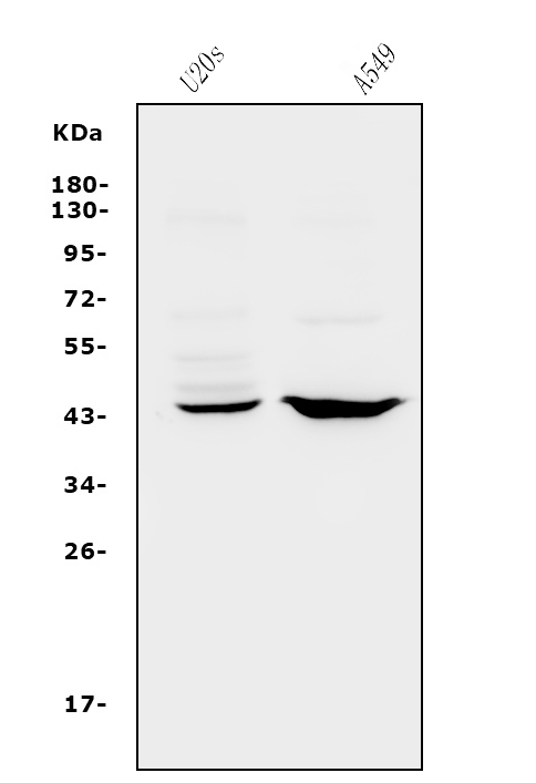
- Main image
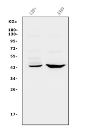
- Experimental details
- Western blot analysis of CXCR2 using anti-CXCR2 antibody (A00455-2). Electrophoresis was performed on a 5-20% SDS-PAGE gel at 70V (Stacking gel) / 90V (Resolving gel) for 2-3 hours. The sample well of each lane was loaded with 30ug of sample under reducing conditions. Lane 1: human U20S whole cell lysates, Lane 2: human A549 whole cell lysates. After Electrophoresis, proteins were transferred to a Nitrocellulose membrane at 150mA for 50-90 minutes. Blocked the membrane with 5% Non-fat Milk/ TBS for 1.5 hour at RT. The membrane was incubated with rabbit anti-CXCR2 antigen affinity purified polyclonal antibody (Catalog # A00455-2) at 0.5 μg/mL overnight at 4°C, then washed with TBS-0.1%Tween 3 times with 5 minutes each and probed with a goat anti-rabbit IgG-HRP secondary antibody at a dilution of 1:5000 for 1.5 hour at RT. The signal is developed using an Enhanced Chemiluminescent detection (ECL) kit (Catalog # EK1002) with Tanon 5200 system. A specific band was detected for CXCR2 at approximately 41KD. The expected band size for CXCR2 is at 41KD.
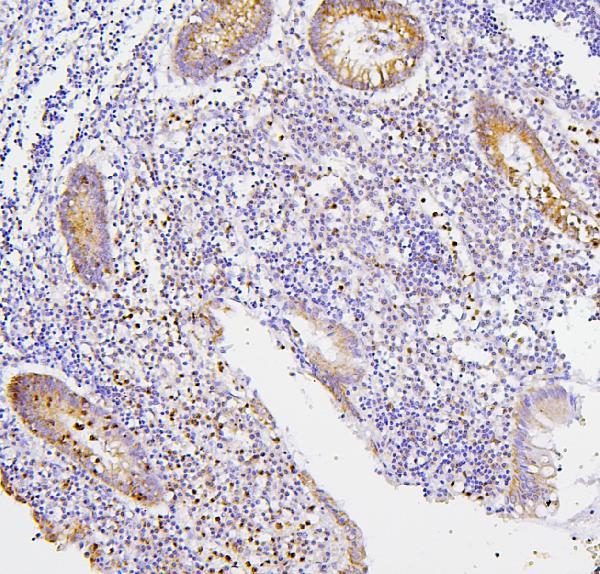
- Additional image