PA1-741
antibody from Invitrogen Antibodies
Targeting: DLG1
dJ1061C18.1.1, DLGH1, hdlg, SAP-97, SAP97
Antibody data
- Antibody Data
- Antigen structure
- References [30]
- Comments [0]
- Validations
- Western blot [2]
- Immunocytochemistry [6]
- Other assay [22]
Submit
Validation data
Reference
Comment
Report error
- Product number
- PA1-741 - Provider product page

- Provider
- Invitrogen Antibodies
- Product name
- SAP97 Polyclonal Antibody
- Antibody type
- Polyclonal
- Antigen
- Synthetic peptide
- Description
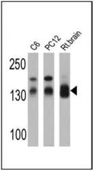
- PA1-741 detects Synapse-Associated Protein 97 (SAP97) from human, rat and mouse tissues. No cross-reactivity to other proteins, including synapse-associated protein family members, has been observed. PA1-741 has been successfully used in Western blot, immunofluorescence, immunocytochemistry, immunoprecipitation, and immunohistochemistry procedures. By Western blot, this antibody detects an ~140 kDa protein representing SAP97 from rat brain extract. Immunohistochemical staining of SAP97 in rat hippocampus with PA1-741 results in intense neuronal cell body and dendrite staining. The PA1-741 immunizing peptide corresponds to amino acids 115-133 from rat SAP97. This sequence is 84% and 74% conserved in mouse and human SAP97, respectively. PA1-741 can be used with blocking peptide PEP-055.
- Reactivity
- Human, Mouse, Rat
- Host
- Rabbit
- Isotype
- IgG
- Vial size
- 100 µL
- Concentration
- Conc. Not Determined
- Storage
- -20° C, Avoid Freeze/Thaw Cycles
Submitted references Quantitative fragmentomics allow affinity mapping of interactomes.
Schizophrenia-associated SAP97 mutations increase glutamatergic synapse strength in the dentate gyrus and impair contextual episodic memory in rats.
SAP97 regulates behavior and expression of schizophrenia risk enriched gene sets in mouse hippocampus.
Proteomic Analysis of Dendritic Filopodia-Rich Fraction Isolated by Telencephalin and Vitronectin Interaction.
Choroid plexus epithelial cells express the adhesion protein P-cadherin at cell-cell contacts and syntaxin-4 in the luminal membrane domain.
SAP97 Binding Partner CRIPT Promotes Dendrite Growth In Vitro and In Vivo.
N-terminal SAP97 isoforms differentially regulate synaptic structure and postsynaptic surface pools of AMPA receptors.
Upregulation of μ3A Drives Homeostatic Plasticity by Rerouting AMPAR into the Recycling Endosomal Pathway.
Dietary and donepezil modulation of mTOR signaling and neuroinflammation in the brain.
Corticotropin-Releasing Hormone Receptor Type 1 (CRHR1) Clustering with MAGUKs Is Mediated via Its C-Terminal PDZ Binding Motif.
Structure-function analysis of SAP97, a modular scaffolding protein that drives dendrite growth.
Differential Changes in Postsynaptic Density Proteins in Postmortem Huntington's Disease and Parkinson's Disease Human Brains.
Sequential delivery of synaptic GluA1- and GluA4-containing AMPA receptors (AMPARs) by SAP97 anchored protein complexes in classical conditioning.
Postsynaptic density scaffold SAP102 regulates cortical synapse development through EphB and PAK signaling pathway.
Adenomatous polyposis coli regulates oligodendroglial development.
Increased [³H]D-aspartate release and changes in glutamate receptor expression in the hippocampus of the mnd mouse.
Cognitive impairment in humanized APP×PS1 mice is linked to Aβ(1-42) and NOX activation.
A specific requirement of Arc/Arg3.1 for visual experience-induced homeostatic synaptic plasticity in mouse primary visual cortex.
Pals1 is a major regulator of the epithelial-like polarization and the extension of the myelin sheath in peripheral nerves.
Subunit- and pathway-specific localization of NMDA receptors and scaffolding proteins at ganglion cell synapses in rat retina.
GluR1 controls dendrite growth through its binding partner, SAP97.
Differential neuronal and glial expression of GluR1 AMPA receptor subunit and the scaffolding proteins SAP97 and 4.1N during rat cerebellar development.
The gap junction protein connexin32 interacts with the Src homology 3/hook domain of discs large homolog 1.
Distribution of the scaffolding proteins PSD-95, PSD-93, and SAP97 in isolated PSDs.
Disruption of Mtmr2 produces CMT4B1-like neuropathy with myelin outfolding and impaired spermatogenesis.
Calcium/calmodulin-dependent protein kinase II phosphorylation drives synapse-associated protein 97 into spines.
Differential recruitment of Kv1.4 and Kv4.2 to lipid rafts by PSD-95.
A multiprotein trafficking complex composed of SAP97, CASK, Veli, and Mint1 is associated with inward rectifier Kir2 potassium channels.
Affinity purification of PSD-95-containing postsynaptic complexes.
Affinity purification of PSD-95-containing postsynaptic complexes.
Gogl G, Zambo B, Kostmann C, Cousido-Siah A, Morlet B, Durbesson F, Negroni L, Eberling P, Jané P, Nominé Y, Zeke A, Østergaard S, Monsellier É, Vincentelli R, Travé G
Nature communications 2022 Sep 17;13(1):5472
Nature communications 2022 Sep 17;13(1):5472
Schizophrenia-associated SAP97 mutations increase glutamatergic synapse strength in the dentate gyrus and impair contextual episodic memory in rats.
Kay Y, Tsan L, Davis EA, Tian C, Décarie-Spain L, Sadybekov A, Pushkin AN, Katritch V, Kanoski SE, Herring BE
Nature communications 2022 Feb 10;13(1):798
Nature communications 2022 Feb 10;13(1):798
SAP97 regulates behavior and expression of schizophrenia risk enriched gene sets in mouse hippocampus.
Gupta P, Uner OE, Nayak S, Grant GR, Kalb RG
PloS one 2018;13(7):e0200477
PloS one 2018;13(7):e0200477
Proteomic Analysis of Dendritic Filopodia-Rich Fraction Isolated by Telencephalin and Vitronectin Interaction.
Furutani Y, Yoshihara Y
Frontiers in synaptic neuroscience 2018;10:27
Frontiers in synaptic neuroscience 2018;10:27
Choroid plexus epithelial cells express the adhesion protein P-cadherin at cell-cell contacts and syntaxin-4 in the luminal membrane domain.
Christensen IB, Mogensen EN, Damkier HH, Praetorius J
American journal of physiology. Cell physiology 2018 May 1;314(5):C519-C533
American journal of physiology. Cell physiology 2018 May 1;314(5):C519-C533
SAP97 Binding Partner CRIPT Promotes Dendrite Growth In Vitro and In Vivo.
Zhang L, Jablonski AM, Mojsilovic-Petrovic J, Ding H, Seeholzer S, Newton IP, Nathke I, Neve R, Zhai J, Shang Y, Zhang M, Kalb RG
eNeuro 2017 Nov-Dec;4(6)
eNeuro 2017 Nov-Dec;4(6)
N-terminal SAP97 isoforms differentially regulate synaptic structure and postsynaptic surface pools of AMPA receptors.
Goodman L, Baddeley D, Ambroziak W, Waites CL, Garner CC, Soeller C, Montgomery JM
Hippocampus 2017 Jun;27(6):668-682
Hippocampus 2017 Jun;27(6):668-682
Upregulation of μ3A Drives Homeostatic Plasticity by Rerouting AMPAR into the Recycling Endosomal Pathway.
Steinmetz CC, Tatavarty V, Sugino K, Shima Y, Joseph A, Lin H, Rutlin M, Lambo M, Hempel CM, Okaty BW, Paradis S, Nelson SB, Turrigiano GG
Cell reports 2016 Sep 6;16(10):2711-2722
Cell reports 2016 Sep 6;16(10):2711-2722
Dietary and donepezil modulation of mTOR signaling and neuroinflammation in the brain.
Dasuri K, Zhang L, Kim SO, Bruce-Keller AJ, Keller JN
Biochimica et biophysica acta 2016 Feb;1862(2):274-83
Biochimica et biophysica acta 2016 Feb;1862(2):274-83
Corticotropin-Releasing Hormone Receptor Type 1 (CRHR1) Clustering with MAGUKs Is Mediated via Its C-Terminal PDZ Binding Motif.
Bender J, Engeholm M, Ederer MS, Breu J, Møller TC, Michalakis S, Rasko T, Wanker EE, Biel M, Martinez KL, Wurst W, Deussing JM
PloS one 2015;10(9):e0136768
PloS one 2015;10(9):e0136768
Structure-function analysis of SAP97, a modular scaffolding protein that drives dendrite growth.
Zhang L, Hsu FC, Mojsilovic-Petrovic J, Jablonski AM, Zhai J, Coulter DA, Kalb RG
Molecular and cellular neurosciences 2015 Mar;65:31-44
Molecular and cellular neurosciences 2015 Mar;65:31-44
Differential Changes in Postsynaptic Density Proteins in Postmortem Huntington's Disease and Parkinson's Disease Human Brains.
Fourie C, Kim E, Waldvogel H, Wong JM, McGregor A, Faull RL, Montgomery JM
Journal of neurodegenerative diseases 2014;2014:938530
Journal of neurodegenerative diseases 2014;2014:938530
Sequential delivery of synaptic GluA1- and GluA4-containing AMPA receptors (AMPARs) by SAP97 anchored protein complexes in classical conditioning.
Zheng Z, Keifer J
The Journal of biological chemistry 2014 Apr 11;289(15):10540-10550
The Journal of biological chemistry 2014 Apr 11;289(15):10540-10550
Postsynaptic density scaffold SAP102 regulates cortical synapse development through EphB and PAK signaling pathway.
Murata Y, Constantine-Paton M
The Journal of neuroscience : the official journal of the Society for Neuroscience 2013 Mar 13;33(11):5040-52
The Journal of neuroscience : the official journal of the Society for Neuroscience 2013 Mar 13;33(11):5040-52
Adenomatous polyposis coli regulates oligodendroglial development.
Lang J, Maeda Y, Bannerman P, Xu J, Horiuchi M, Pleasure D, Guo F
The Journal of neuroscience : the official journal of the Society for Neuroscience 2013 Feb 13;33(7):3113-30
The Journal of neuroscience : the official journal of the Society for Neuroscience 2013 Feb 13;33(7):3113-30
Increased [³H]D-aspartate release and changes in glutamate receptor expression in the hippocampus of the mnd mouse.
Bigini P, Milanese M, Gardoni F, Longhi A, Bonifacino T, Barbera S, Fumagalli E, Di Luca M, Mennini T, Bonanno G
Journal of neuroscience research 2012 Jun;90(6):1148-58
Journal of neuroscience research 2012 Jun;90(6):1148-58
Cognitive impairment in humanized APP×PS1 mice is linked to Aβ(1-42) and NOX activation.
Bruce-Keller AJ, Gupta S, Knight AG, Beckett TL, McMullen JM, Davis PR, Murphy MP, Van Eldik LJ, St Clair D, Keller JN
Neurobiology of disease 2011 Dec;44(3):317-26
Neurobiology of disease 2011 Dec;44(3):317-26
A specific requirement of Arc/Arg3.1 for visual experience-induced homeostatic synaptic plasticity in mouse primary visual cortex.
Gao M, Sossa K, Song L, Errington L, Cummings L, Hwang H, Kuhl D, Worley P, Lee HK
The Journal of neuroscience : the official journal of the Society for Neuroscience 2010 May 26;30(21):7168-78
The Journal of neuroscience : the official journal of the Society for Neuroscience 2010 May 26;30(21):7168-78
Pals1 is a major regulator of the epithelial-like polarization and the extension of the myelin sheath in peripheral nerves.
Ozçelik M, Cotter L, Jacob C, Pereira JA, Relvas JB, Suter U, Tricaud N
The Journal of neuroscience : the official journal of the Society for Neuroscience 2010 Mar 17;30(11):4120-31
The Journal of neuroscience : the official journal of the Society for Neuroscience 2010 Mar 17;30(11):4120-31
Subunit- and pathway-specific localization of NMDA receptors and scaffolding proteins at ganglion cell synapses in rat retina.
Zhang J, Diamond JS
The Journal of neuroscience : the official journal of the Society for Neuroscience 2009 Apr 1;29(13):4274-86
The Journal of neuroscience : the official journal of the Society for Neuroscience 2009 Apr 1;29(13):4274-86
GluR1 controls dendrite growth through its binding partner, SAP97.
Zhou W, Zhang L, Guoxiang X, Mojsilovic-Petrovic J, Takamaya K, Sattler R, Huganir R, Kalb R
The Journal of neuroscience : the official journal of the Society for Neuroscience 2008 Oct 8;28(41):10220-33
The Journal of neuroscience : the official journal of the Society for Neuroscience 2008 Oct 8;28(41):10220-33
Differential neuronal and glial expression of GluR1 AMPA receptor subunit and the scaffolding proteins SAP97 and 4.1N during rat cerebellar development.
Douyard J, Shen L, Huganir RL, Rubio ME
The Journal of comparative neurology 2007 May 1;502(1):141-56
The Journal of comparative neurology 2007 May 1;502(1):141-56
The gap junction protein connexin32 interacts with the Src homology 3/hook domain of discs large homolog 1.
Duffy HS, Iacobas I, Hotchkiss K, Hirst-Jensen BJ, Bosco A, Dandachi N, Dermietzel R, Sorgen PL, Spray DC
The Journal of biological chemistry 2007 Mar 30;282(13):9789-96
The Journal of biological chemistry 2007 Mar 30;282(13):9789-96
Distribution of the scaffolding proteins PSD-95, PSD-93, and SAP97 in isolated PSDs.
DeGiorgis JA, Galbraith JA, Dosemeci A, Chen X, Reese TS
Brain cell biology 2006 Dec;35(4-6):239-50
Brain cell biology 2006 Dec;35(4-6):239-50
Disruption of Mtmr2 produces CMT4B1-like neuropathy with myelin outfolding and impaired spermatogenesis.
Bolino A, Bolis A, Previtali SC, Dina G, Bussini S, Dati G, Amadio S, Del Carro U, Mruk DD, Feltri ML, Cheng CY, Quattrini A, Wrabetz L
The Journal of cell biology 2004 Nov 22;167(4):711-21
The Journal of cell biology 2004 Nov 22;167(4):711-21
Calcium/calmodulin-dependent protein kinase II phosphorylation drives synapse-associated protein 97 into spines.
Mauceri D, Cattabeni F, Di Luca M, Gardoni F
The Journal of biological chemistry 2004 May 28;279(22):23813-21
The Journal of biological chemistry 2004 May 28;279(22):23813-21
Differential recruitment of Kv1.4 and Kv4.2 to lipid rafts by PSD-95.
Wong W, Schlichter LC
The Journal of biological chemistry 2004 Jan 2;279(1):444-52
The Journal of biological chemistry 2004 Jan 2;279(1):444-52
A multiprotein trafficking complex composed of SAP97, CASK, Veli, and Mint1 is associated with inward rectifier Kir2 potassium channels.
Leonoudakis D, Conti LR, Radeke CM, McGuire LM, Vandenberg CA
The Journal of biological chemistry 2004 Apr 30;279(18):19051-63
The Journal of biological chemistry 2004 Apr 30;279(18):19051-63
Affinity purification of PSD-95-containing postsynaptic complexes.
Vinade L, Chang M, Schlief ML, Petersen JD, Reese TS, Tao-Cheng JH, Dosemeci A
Journal of neurochemistry 2003 Dec;87(5):1255-61
Journal of neurochemistry 2003 Dec;87(5):1255-61
Affinity purification of PSD-95-containing postsynaptic complexes.
Vinade L, Chang M, Schlief ML, Petersen JD, Reese TS, Tao-Cheng JH, Dosemeci A
Journal of neurochemistry 2003 Dec;87(5):1255-61
Journal of neurochemistry 2003 Dec;87(5):1255-61
No comments: Submit comment
Supportive validation
- Submitted by
- Invitrogen Antibodies (provider)
- Main image
- Experimental details
- Western blot analysis of SAP97 was performed by loading 25 µg of C6 (lane 1), PC12 (lane 2) and rat brain (lane 3) cell lysates onto an SDS polyacrylamide gel. Proteins were transferred to a PVDF membrane and blocked at 4ºC overnight. The membrane was probed with a SAP97 polyclonal antibody (Product # PA1-741) at a dilution of 1:5000 overnight at 4°C, washed in TBST, and probed with an HRP-conjugated secondary antibody for 1 hr at room temperature in the dark. Chemiluminescent detection was performed using Pierce ECL Plus Western Blotting Substrate (Product # 32132). Results show a band at ~140 kDa.
- Submitted by
- Invitrogen Antibodies (provider)
- Main image
- Experimental details
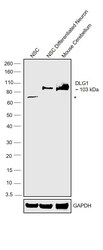
- Western blot was performed using Anti-SAP97 Polyclonal Antibody (Product # PA1-741) and a 103 kDa band corresponding to DLG1 was observed in neural stem cells (NSC) differentiated into neurons and Mouse Cerebellum along with an uncharacterized band (*) was NSC. Whole cell extracts (40 µg lysate) of NSC (Lane 1), NSC differentiated into neurons (Lane 2), and tissue extract (40 µg lysate) of Mouse Cerebellum (Lane 3) were electrophoresed using NuPAGE™ 10% Bis-Tris Protein Gel (Product # NP0301BOX). Resolved proteins were then transferred onto a nitrocellulose membrane (Product # IB23001) by iBlot® 2 Dry Blotting System (Product # IB21001). The blot was probed with the primary antibody (1:1000) and detected by chemiluminescence with Goat anti-Rabbit IgG (H+L) Superclonal™ Recombinant Secondary Antibody, HRP (Product # A27036,1:4000) using the iBright™ FL1500 Imaging System (Product # A44115). Chemiluminescentdetection was performed using Novex® ECL Chemiluminescent Substrate Reagent Kit (Product # WP20005).
Supportive validation
- Submitted by
- Invitrogen Antibodies (provider)
- Main image
- Experimental details
- Immunofluorescent analysis of SAP97 (green) showing staining in the cytoplasm and membrane of C2C12 cells. Formalin-fixed cells were permeabilized with 0.1% Triton X-100 in TBS for 5-10 minutes and blocked with 3% BSA-PBS for 30 minutes at room temperature. Cells were probed with a SAP97 polyclonal antibody (Product # PA1-741) in 3% BSA-PBS at a dilution of 1:100 and incubated overnight at 4 ºC in a humidified chamber. Cells were washed with PBST and incubated with a DyLight-conjugated secondary antibody in PBS at room temperature in the dark. F-actin (red) was stained with a fluorescent red phalloidin and nuclei (blue) were stained with Hoechst or DAPI. Images were taken at a magnification of 60x.
- Submitted by
- Invitrogen Antibodies (provider)
- Main image

- Experimental details
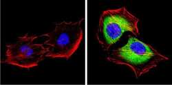
- Immunofluorescent analysis of SAP97 (green) showing staining in the cytoplasm and membrane of HeLa cells. Formalin-fixed cells were permeabilized with 0.1% Triton X-100 in TBS for 5-10 minutes and blocked with 3% BSA-PBS for 30 minutes at room temperature. Cells were probed with a SAP97 polyclonal antibody (Product # PA1-741) in 3% BSA-PBS at a dilution of 1:100 and incubated overnight at 4 ºC in a humidified chamber. Cells were washed with PBST and incubated with a DyLight-conjugated secondary antibody in PBS at room temperature in the dark. F-actin (red) was stained with a fluorescent red phalloidin and nuclei (blue) were stained with Hoechst or DAPI. Images were taken at a magnification of 60x.
- Submitted by
- Invitrogen Antibodies (provider)
- Main image
- Experimental details
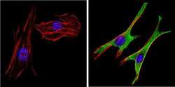
- Immunofluorescent analysis of SAP97 (green) showing staining in the cytoplasm and membrane of NIH-3T3 cells. Formalin-fixed cells were permeabilized with 0.1% Triton X-100 in TBS for 5-10 minutes and blocked with 3% BSA-PBS for 30 minutes at room temperature. Cells were probed with a SAP97 polyclonal antibody (Product # PA1-741) in 3% BSA-PBS at a dilution of 1:100 and incubated overnight at 4 ºC in a humidified chamber. Cells were washed with PBST and incubated with a DyLight-conjugated secondary antibody in PBS at room temperature in the dark. F-actin (red) was stained with a fluorescent red phalloidin and nuclei (blue) were stained with Hoechst or DAPI. Images were taken at a magnification of 60x.
- Submitted by
- Invitrogen Antibodies (provider)
- Main image
- Experimental details
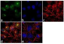
- Immunofluorescence analysis of SAP97 was performed using 70% confluent log phase RSC96 cells. The cells were fixed with 4% paraformaldehyde for 10 minutes, permeabilized with 0.1% Triton™ X-100 for 10 minutes, and blocked with 1% BSA for 1 hour at room temperature. The cells were labeled with SAP97 Rabbit Polyclonal Antibody (Product # PA1-741) at 1:250 dilution in 0.1% BSA and incubated for 3 hours at room temperature and then labeled with Goat anti-Rabbit IgG (H+L) Superclonal™ Secondary Antibody, Alexa Fluor® 488 conjugate (Product # A27034) at a dilution of 1:2000 for 45 minutes at room temperature (Panel a: green). Nuclei (Panel b: blue) were stained with SlowFade® Gold Antifade Mountant with DAPI (Product # S36938). F-actin (Panel c: red) was stained with Rhodamine Phalloidin (Product # R415, 1:300). Panel d represents the merged image showing cytoplasmic localization. Panel e shows the no primary antibody control. The images were captured at 60X magnification.
- Submitted by
- Invitrogen Antibodies (provider)
- Main image
- Experimental details
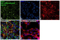
- Immunofluorescence analysis of DLG1 was performed using neural stem cells (NSC) differentiated into neurons. The cells were fixed with 4% paraformaldehyde for 10 minutes, permeabilized with 0.1% Triton™ X-100 for 10 minutes, and blocked with 2% BSA for 45 minutes at room temperature. The cells were labeled with SAP97 Polyclonal Antibody (Product # PA1-741) at 1:200 in 0.1% BSA, incubated at 4 degree celsius overnight and then labeled with Goat anti-Rabbit IgG (H+L) Superclonal™ Recombinant Secondary Antibody, Alexa Fluor® 488 conjugate (Product # A27034), (1:2500), for 45 minutes at room temperature (Panel a: Green). Nuclei (Panel b:Blue) were stained with ProLong™ Diamond Antifade Mountant with DAPI (Product # P36962). F-actin (Panel c: Red) was stained with Rhodamine Phalloidin (Product # R415, 1:300). Panel d represents the merged image showing plasma membrane and synaptic junctional localization. Panel e represents control cells with no primary antibody to assess background. The images were captured at 60X magnification.
- Submitted by
- Invitrogen Antibodies (provider)
- Main image
- Experimental details
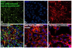
- Immunofluorescence analysis of DLG1 was performed using neural stem cells (NSC) differentiated into neurons. The cells were fixed with 4% paraformaldehyde for 10 minutes, permeabilized with 0.1% Triton™ X-100 for 10 minutes, and blocked with 2% BSA for 45 minutes at room temperature. The cells were labeled with SAP97 Polyclonal Antibody (Product # PA1-741) at 1:200 in 0.1% BSA, incubated at 4 degree celsius overnight and then labeled with Goat anti-Rabbit IgG (H+L) Superclonal™ Recombinant Secondary Antibody, Alexa Fluor® 488 conjugate (Product # A27034), (1:2500), for 45 minutes at room temperature (Panel a: Green). Nuclei (Panel b: Blue) were stained with ProLong™ Diamond Antifade Mountant with DAPI (Product # P36962). F-actin (Panel c: Red) was stained with Rhodamine Phalloidin (Product # R415, 1:300). Panel d represents the merged image showing plasma membrane and synaptic junctional localization. Panel e represents low level expression in undifferentiated NSCs. Panel f represents control cells with no primary antibody to assess background. The images were captured at 60X magnification.
Supportive validation
- Submitted by
- Invitrogen Antibodies (provider)
- Main image
- Experimental details
- NULL
- Submitted by
- Invitrogen Antibodies (provider)
- Main image
- Experimental details
- NULL
- Submitted by
- Invitrogen Antibodies (provider)
- Main image
- Experimental details
- NULL
- Submitted by
- Invitrogen Antibodies (provider)
- Main image
- Experimental details
- NULL
- Submitted by
- Invitrogen Antibodies (provider)
- Main image
- Experimental details
- NULL
- Submitted by
- Invitrogen Antibodies (provider)
- Main image
- Experimental details
- NULL
- Submitted by
- Invitrogen Antibodies (provider)
- Main image
- Experimental details
- NULL
- Submitted by
- Invitrogen Antibodies (provider)
- Main image
- Experimental details
- NULL
- Submitted by
- Invitrogen Antibodies (provider)
- Main image
- Experimental details
- NULL
- Submitted by
- Invitrogen Antibodies (provider)
- Main image
- Experimental details
- NULL
- Submitted by
- Invitrogen Antibodies (provider)
- Main image
- Experimental details
- NULL
- Submitted by
- Invitrogen Antibodies (provider)
- Main image

- Experimental details
- NULL
- Submitted by
- Invitrogen Antibodies (provider)
- Main image
- Experimental details
- NULL
- Submitted by
- Invitrogen Antibodies (provider)
- Main image

- Experimental details
- NULL
- Submitted by
- Invitrogen Antibodies (provider)
- Main image

- Experimental details
- NULL
- Submitted by
- Invitrogen Antibodies (provider)
- Main image

- Experimental details
- NULL
- Submitted by
- Invitrogen Antibodies (provider)
- Main image
- Experimental details
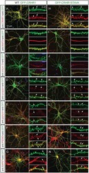
- Fig 7 CRHR1-WT and CRHR1-STAVA localize throughout the neuronal plasma membrane including the excitatory post synapse. Primary hippocampal neurons at DIV 14-20 were transduced with CRHR1-WT and CRHR1-STAVA using AAV8. (A) The spatial pattern of CRHR1-WT and CRHR1-STAVA expression visualized by immunostaining of the GFP tag was confirmed using an anti-CRHR1 antibody. (B) Microtubule-associated protein 2 (MAP2) staining revealed the dendritic presence of the receptor. (C) Co-staining with the axon initial segment marker ankyrin G demonstrated CRHR1-WT and CRHR1-STAVA localization within axons. (D) The presynaptic marker synapsin indicated that CRHR1-WT and CRHR1-STAVA are present in the adjacent post synapse but not in the presynaptic axon terminal. (E) CRHR1-WT and CRHR1-STAVA did not co-localize with the inhibitory postsynaptic marker gephyrin but (F) they co-localized with the MAGUK and excitatory postsynaptic marker PSD95 in spines. (G) CRHR1-WT and mutant co-localized with the candidate interaction partner SAP97.
- Submitted by
- Invitrogen Antibodies (provider)
- Main image
- Experimental details
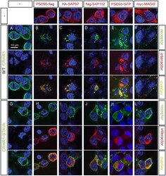
- Fig 8 CRHR1 PDZ binding motif is required for clustering with interacting MAGUKs. CRHR1-WT (A), CRHR1-STAVA (G), or the interacting proteins (first row) were transiently transfected alone. (B-F) Wild-type CRHR1 or (H-L) CRHR1-STAVA were transiently transfected in HEK293 together with (B, H) PSD95-flag, (C, I) HA-SAP97, (D, J) flag-SAP102, (E, K) PSD93-GFP and (F, L) myc-MAGI2. CRHR1-WT co-transfection with the respective interacting partner resulted in a co-clustering of both proteins (B-F). (H-L) Co-transfection of CRHR1-STAVA with the respective interacting partner did not result in any clustering, but both proteins appeared with a subcellular distribution similar to the individual transfections. Immunostaining was performed against the HA tag of HA-CRHR1 or HA-CRHR1-STAVA when co-transfected with PSD95-flag or flag-SAP102. Immunostaining was performed against the flag tag of flag-CRHR1 or flag-CRHR1-STAVA when co-transfected with PSD93-GFP, HA-SAP97, or myc-MAGI2 respectively.
- Submitted by
- Invitrogen Antibodies (provider)
- Main image
- Experimental details
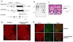
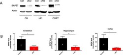
- Fig 1 SAP97 protein is sufficiently knocked down in SAP97-cKO animals. (A) Western blots showing reduced SAP97 band intensity in cerebellum, hippocampus, and cortex. (B) Quantification of western blot analysis. *P < .05, **P < .01 (two-tailed Student's t test). Data are presented as mean +- SEM.
- Submitted by
- Invitrogen Antibodies (provider)
- Main image
- Experimental details
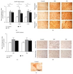
- Figure 2 SAP97 expression in human HD and PD hippocampus and striatum. (a) Significant increases in SAP97 expression in the post-mortem human hippocampus in HD and PD patients. The significant increase in SAP97 expression occurs in all hippocampal regions examined: DG, area CA3, and area CA1. * P < 0.05, ** P < 0.005. (b) No significant changes in SAP97 expression were observed in HD or PD striatum in either the caudate nucleus (CN) or the putamen (Put). (c) Representative images of SAP97 immunostaining in the dentate gyrus and CA3 and CA1 hippocampal regions. (d) Representative striatal images of SAP97 immunostaining in the putamen and caudate nucleus. Scale bar 25 mu m for (c) and (d). (e) High power example image of SAP97 immunolabeling, showing the expected somatic and dendritic localisations.
- Submitted by
- Invitrogen Antibodies (provider)
- Main image
- Experimental details
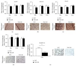
- Figure 5 Quantitative immunohistochemistry of SAP97, PSD-95, GluN1, and GluA2 expression in YAC128 hippocampal sections. Sections were prepared from symptomatic 1-year-old YAC128 mice to provide a comparison to the end stage of human HD. (a)-(d). Top: Quantification of (a) SAP97, (b) PSD-95, (c) GluA2, and (d) GluN1 levels in dentate gyrus (DG), area CA3, and area CA1. Below: Example immunohistochemical staining for each glutamatergic synaptic protein in the hippocampal CA1 region in control (wildtype) and YAC128 mice. (e) Immunohistochemical quantification of DARPP-32 expression in wild-type and YAC128 striatum (caudate putamen, CPu). * P < 0.05.
- Submitted by
- Invitrogen Antibodies (provider)
- Main image
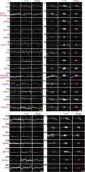
- Experimental details
- FIGURE 4 Validation of the identified proteins in dendritic filopodia. (A,B) Localization of the identified proteins in dendritic filopodia (A1-A21) and phagocytic cups (B1-B21) at 14 DIV hippocampal neurons. The cultured neurons were immunostained with anti-TLCN antibody and specific antibodies against Galphao (A1,B1) , Na + /K + ATPase alpha3 (A2,B2) , EFA6D (A3,B3 ), spectrin (A4,B4) , septin7 (A5,B5) , myosin VA (A6,B6) , Galphaq (A7,B7) , Gbeta2 (A8,B8) , eEF1gamma (A9,B9) , ribosomal protein S16 (A10,B10) , CaMKIIalpha (A11,B11) , CD98hc (A12,B12) , EFA6C (A13,B13) , BAIAP2L1 (A14,B14) , PLCbeta3 (A15,B15) , MRCKalpha (A16,B16) , NR3A/B (GluN3A/B) (A17,B17) , SAP97 (SLC3A2) (A18,B18) , MAP1S (A19,B19) , EPS8L1 (A20,B20) , and JIP-4 (SPAG9) (A21,B21) . In merged images, the identified proteins and TLCN are shown in magenta and green, respectively. Single plane of images focused on dendritic filopodia (A1-A21) and center of microbeads (B1-B21) were acquired using a confocal microscopy. Scale bars, 5 mum in (A14,A21,B14,B21) .
 Explore
 Validate
 Learn
Learn Western blot
 Immunoprecipitation