Explore
 Validate
 Learn
Learn Western blot
Antibody data
- Antibody Data
- Antigen structure
- References [0]
- Comments [0]
- Validations
- Western blot [2]
- Immunohistochemistry [4]
Submit
Validation data
Reference
Comment
Report error
- Product number
- AP42051PU-N - Provider product page

- Provider
- Acris Antibodies GmbH
- Proper citation
- Acris Antibodies GmbH Cat#AP42051PU-N, RRID:AB_11216062
- Product name
- anti KIN
- Antibody type
- Polyclonal
- Antigen
- The immunogen for anti-KIN antibody: synthetic peptide directed towards the middle region of human KIN.
- Reactivity
- Human, Bovine, Canine
- Host
- Rabbit
- Vial size
- 50 µg
No comments: Submit comment
Supportive validation
- Submitted by
- Acris Antibodies GmbH (provider)
- Main image
- Experimental details
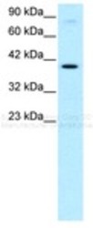
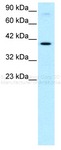
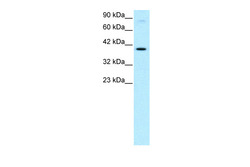
- Western blotting of human HepG2 cell lysate using AP42051PU-N KIN antibody
- Submitted by
- Acris Antibodies GmbH (provider)
- Main image
- Experimental details
- Human HepG2; WB Suggested Anti-KIN Antibody Titration: 0.2-1 ug/ml. ELISA Titer: 1:2500. Positive Control: HepG2 cell lysate. KIN is supported by BioGPS gene expression data to be expressed in HepG2.; KIN antibody - middle region (AP42051PU-N) in Human HepG2 cells using Western Blot

Supportive validation
- Submitted by
- Acris Antibodies GmbH (provider)
- Main image
- Experimental details
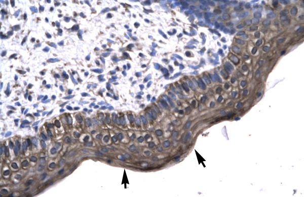
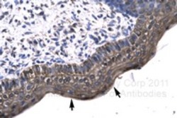
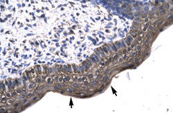
- Staining of paraffin embedded sections of human spermatophore using AP42051PU-N KIN antibody. Note positive staining of epithelial cells (arrows).
- Submitted by
- Acris Antibodies GmbH (provider)
- Main image
- Experimental details
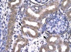
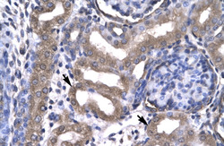
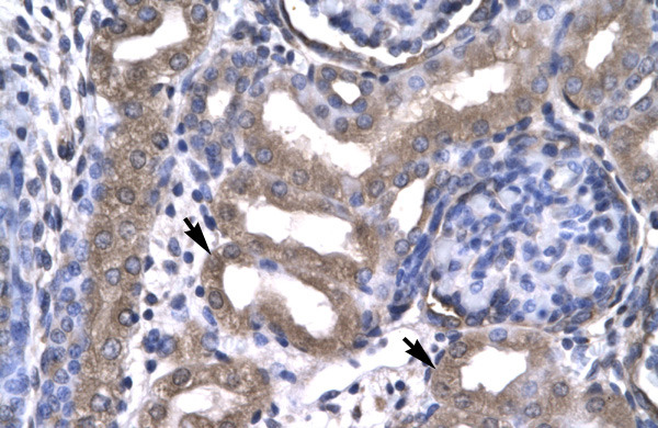
- Staining of paraffin embedded sections of human kidney using AP42051PU-N KIN antibody. Note positive staining of epithelial cells of renal tubule (arrows).
- Submitted by
- Acris Antibodies GmbH (provider)
- Main image
- Experimental details
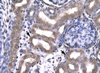
- Human kidney; KIN antibody - middle region (AP42051PU-N) in Human kidney cells using Immunohistochemistry
- Submitted by
- Acris Antibodies GmbH (provider)
- Main image
- Experimental details
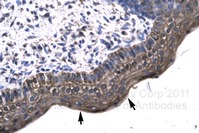
- Human Spermatophore; KIN antibody - middle region (AP42051PU-N) in Human Spermatophore cells using Immunohistochemistry