Explore
 Validate
 Learn
LearnPA5-114344
antibody from Invitrogen Antibodies
Targeting: CXCL12
PBSF, SCYB12, SDF-1a, SDF-1b, SDF1, SDF1A, SDF1B, TLSF-a, TLSF-b, TPAR1
 Western blot
Antibody data
- Antibody Data
- Antigen structure
- References [3]
- Comments [0]
- Validations
- Western blot [1]
- Immunocytochemistry [2]
- Immunohistochemistry [4]
- Flow cytometry [1]
- Other assay [2]
Submit
Validation data
Reference
Comment
Report error
- Product number
- PA5-114344 - Provider product page

- Provider
- Invitrogen Antibodies
- Product name
- SDF1 Polyclonal Antibody
- Antibody type
- Polyclonal
- Antigen
- Recombinant full-length protein
- Reactivity
- Human, Mouse, Rat
- Host
- Rabbit
- Isotype
- IgG
- Vial size
- 100 µL
- Concentration
- 1 mg/mL
- Storage
- Store at 4°C short term. For long term storage, store at -20°C, avoiding freeze/thaw cycles.
Submitted references Use of gliptins reduces levels of SDF-1/CXCL12 in bullous pemphigoid and type 2 diabetes, but does not increase autoantibodies against BP180 in diabetic patients.
The SDF1-CXCR4 Axis Is Involved in the Hyperbaric Oxygen Therapy-Mediated Neuronal Cells Migration in Transient Brain Ischemic Rats.
Single Cell Analysis of Cultivated Fibroblasts from Chronic Pancreatitis and Pancreatic Cancer Patients.
Nätynki A, Leisti P, Tuusa J, Varpuluoma O, Huilaja L, Izumi K, Herukka SK, Ukkola O, Junttila J, Kokkonen N, Tasanen K
Frontiers in immunology 2022;13:942131
Frontiers in immunology 2022;13:942131
The SDF1-CXCR4 Axis Is Involved in the Hyperbaric Oxygen Therapy-Mediated Neuronal Cells Migration in Transient Brain Ischemic Rats.
Wang RY, Yang YR, Chang HC
International journal of molecular sciences 2022 Feb 4;23(3)
International journal of molecular sciences 2022 Feb 4;23(3)
Single Cell Analysis of Cultivated Fibroblasts from Chronic Pancreatitis and Pancreatic Cancer Patients.
Sunami Y, Chen Y, Trojanowicz B, Sommerer M, Hämmerle M, Eils R, Kleeff J
Cells 2022 Aug 19;11(16)
Cells 2022 Aug 19;11(16)
No comments: Submit comment
Supportive validation
- Submitted by
- Invitrogen Antibodies (provider)
- Main image

- Experimental details
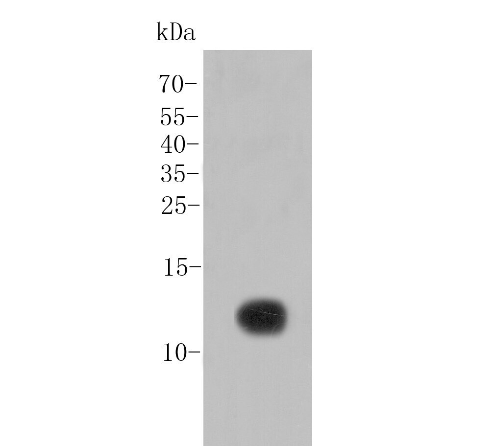
- Western blot analysis of SDF1 using a SDF1 polyclonal antibody (Product #PA5-114344). Proteins were transferred to a PVDF membrane and blocked with 5% BSA in PBS for 1 hour at room temperature. The primary antibody at a dilution of 1:20,000 was used in 5% BSA at room temperature for 2 hours. Goat Anti-Rabbit IgG - HRP Secondary Antibody at 1:5,000 dilution was used for 1 hour at room temperature.
Supportive validation
- Submitted by
- Invitrogen Antibodies (provider)
- Main image
- Experimental details
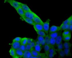
- Immunocytochemical analysis of SDF1 in 293T cells using a SDF1 polyclonal antibody (Product #PA5-114344). Formalin fixed cells were permeabilized with 0.1% Triton X-100 in TBS for 10 minutes at room temperature and blocked with 1% Blocker BSA for 15 minutes at room temperature. Cells were probed with the primary antibody for 1 hour at room temperature, washed with PBS. Alexa Fluor®488 Goat anti-Rabbit IgG was used as the secondary antibody at 1:100 dilution. The nuclear counter stain is DAPI.

- Submitted by
- Invitrogen Antibodies (provider)
- Main image
- Experimental details
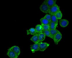
- Immunocytochemical analysis of SDF1 in LOVO cells using a SDF1 polyclonal antibody (Product #PA5-114344). Formalin fixed cells were permeabilized with 0.1% Triton X-100 in TBS for 10 minutes at room temperature and blocked with 1% Blocker BSA for 15 minutes at room temperature. Cells were probed with the primary antibody for 1 hour at room temperature, washed with PBS. Alexa Fluor®488 Goat anti-Rabbit IgG was used as the secondary antibody at 1:100 dilution. The nuclear counter stain is DAPI.

Supportive validation
- Submitted by
- Invitrogen Antibodies (provider)
- Main image
- Experimental details
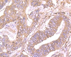
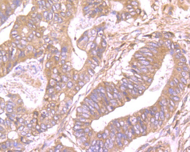
- Immunohistochemical analysis of SDF1 in paraffin-embedded human colon cancer tissue using a polyclonal antibody (Product #PA5-114344). The section was pre-treated using heat mediated antigen retrieval with Tris-EDTA buffer (pH 8.0-8.4) for 20 minutes. The tissues were blocked in 5% BSA for 30 minutes at room temperature, washed with ddH2O and PBS, and then probed with the primary antibody (1:100) for 30 minutes at room temperature. The detection was performed using an HRP conjugated compact polymer system. DAB was used as the chromogen. Tissues were counterstained with hematoxylin and mounted with DPX.
- Submitted by
- Invitrogen Antibodies (provider)
- Main image
- Experimental details
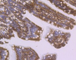
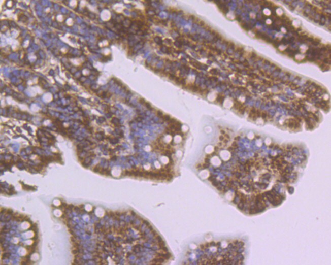
- Immunohistochemical analysis of SDF1 in paraffin-embedded mouse colon tissue using a polyclonal antibody (Product #PA5-114344). The section was pre-treated using heat mediated antigen retrieval with Tris-EDTA buffer (pH 8.0-8.4) for 20 minutes. The tissues were blocked in 5% BSA for 30 minutes at room temperature, washed with ddH2O and PBS, and then probed with the primary antibody (1:100) for 30 minutes at room temperature. The detection was performed using an HRP conjugated compact polymer system. DAB was used as the chromogen. Tissues were counterstained with hematoxylin and mounted with DPX.
- Submitted by
- Invitrogen Antibodies (provider)
- Main image

- Experimental details
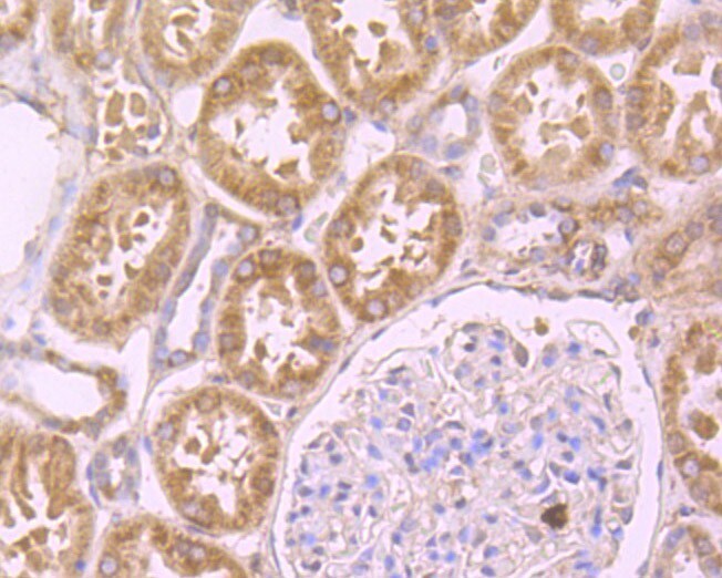
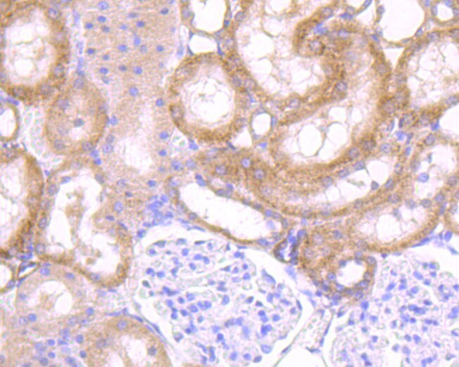
- Immunohistochemical analysis of SDF1 in paraffin-embedded human kidney tissue using a polyclonal antibody (Product #PA5-114344). The section was pre-treated using heat mediated antigen retrieval with Tris-EDTA buffer (pH 8.0-8.4) for 20 minutes. The tissues were blocked in 5% BSA for 30 minutes at room temperature, washed with ddH2O and PBS, and then probed with the primary antibody (1:100) for 30 minutes at room temperature. The detection was performed using an HRP conjugated compact polymer system. DAB was used as the chromogen. Tissues were counterstained with hematoxylin and mounted with DPX.
- Submitted by
- Invitrogen Antibodies (provider)
- Main image
- Experimental details
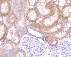
- Immunohistochemical analysis of SDF1 in paraffin-embedded Rat kidney tissue using a polyclonal antibody (Product #PA5-114344). The section was pre-treated using heat mediated antigen retrieval with Tris-EDTA buffer (pH 8.0-8.4) for 20 minutes. The tissues were blocked in 5% BSA for 30 minutes at room temperature, washed with ddH2O and PBS, and then probed with the primary antibody (1:100) for 30 minutes at room temperature. The detection was performed using an HRP conjugated compact polymer system. DAB was used as the chromogen. Tissues were counterstained with hematoxylin and mounted with DPX.
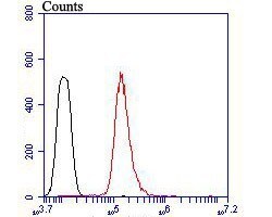
Supportive validation
- Submitted by
- Invitrogen Antibodies (provider)
- Main image
- Experimental details
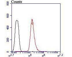
- Flow cytometric analysis of SDF1 using HepG2 cells and a SDF1 polyclonal antibody (Product #PA5-114344). The cells were fixed, permeabilized and stained with the primary antibody at a dilution of 1:100 (red). After incubation of the primary antibody at room temperature for an hour, the cells were stained with a Alexa Fluor 488-conjugated goat anti-rabbit IgG Secondary antibody at 1:500 dilution for 30 minutes. Unlabeled sample was used as a control (cells without incubation with primary antibody; black).
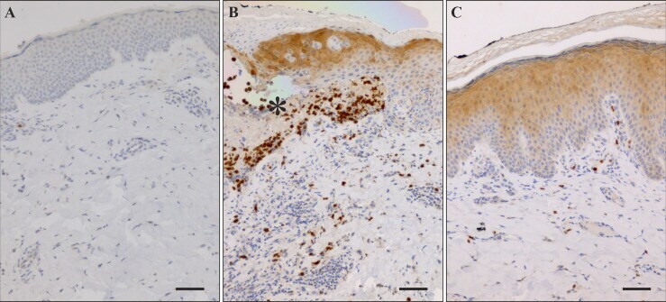
Supportive validation
- Submitted by
- Invitrogen Antibodies (provider)
- Main image

- Experimental details
- Immunostaining of SDF-1 in healthy control skin and lesional BP skin. (A) Immunohistochemical staining of SDF-1 was negative in healthy control skin. (B) Numerous SDF-1 positive cells were detected near blisters and in blister fluid (asterisk) in lesional BP skin along with strong staining of epidermis on the blister roof and on the margins of the blister. (C) In BP perilesional skin sections without blister there is strong staining of the upper layers of the epidermis and relatively few positive cells in the dermis. Scale bar = 50 um.
- Submitted by
- Invitrogen Antibodies (provider)
- Main image
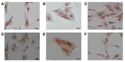
- Experimental details
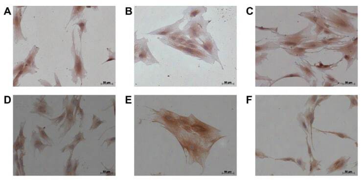
- Immunocytochemistry of alphaSMA and CXCL12 in disease-associated fibroblasts from pancreatic cancer and chronic pancreatitis patients. Cells from ( A ) patient1, ( B ) from patient2, and ( C ) from patient3 for alphaSMA staining, ( D ) cells from patient1, ( E ) from patient2, and ( F ) from patient3 for CXCL12 staining. Bar: 50 mum.