Explore
 Validate
 Learn
Learn Western blot
Antibody data
- Antibody Data
- Antigen structure
- References [1]
- Comments [0]
- Validations
- Western blot [4]
- Immunocytochemistry [1]
- Other assay [2]
Submit
Validation data
Reference
Comment
Report error
- Product number
- PA5-29927 - Provider product page

- Provider
- Invitrogen Antibodies
- Product name
- CREM Polyclonal Antibody
- Antibody type
- Polyclonal
- Antigen
- Recombinant protein fragment
- Description
- Recommended positive controls: NT2D1, Neuro2A, GL261, PC-12, Rat2.
- Concentration
- 0.31 mg/mL
Submitted references Impact of the acidic environment on gene expression and functional parameters of tumors in vitro and in vivo.
Rauschner M, Lange L, Hüsing T, Reime S, Nolze A, Maschek M, Thews O, Riemann A
Journal of experimental & clinical cancer research : CR 2021 Jan 6;40(1):10
Journal of experimental & clinical cancer research : CR 2021 Jan 6;40(1):10
No comments: Submit comment
Supportive validation
- Submitted by
- Invitrogen Antibodies (provider)
- Main image

- Experimental details
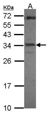
- Western Blot using CREM Polyclonal Antibody (Product # PA5-29927). Sample (30 µg of whole cell lysate). Lane A: NT2D1. 12% SDS PAGE. CREM Polyclonal Antibody . CREM Polyclonal Antibody (Product # PA5-29927) diluted at 1:1,000.
- Submitted by
- Invitrogen Antibodies (provider)
- Main image
- Experimental details
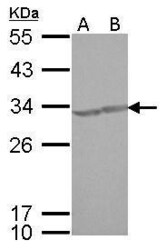
- CREM Polyclonal Antibody detects CREM protein by Western blot analysis. A. 30 µg Neuro2A whole cell lysate/extract. B. 30 µg GL261 whole cell lysate/extract.12 % SDS-PAGE. CREM Polyclonal Antibody (Product # PA5-29927) dilution: 1:1,000.

- Submitted by
- Invitrogen Antibodies (provider)
- Main image

- Experimental details
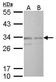
- CREM Polyclonal Antibody detects CREM protein by Western blot analysis. A. 30 µg PC-12 whole cell lysate/extract. B. 30 µg Rat2 whole cell lysate/extract.12 % SDS-PAGE. CREM Polyclonal Antibody (Product # PA5-29927) dilution: 1:1,000.
- Submitted by
- Invitrogen Antibodies (provider)
- Main image
- Experimental details
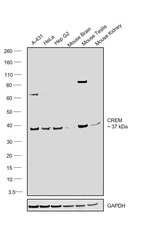
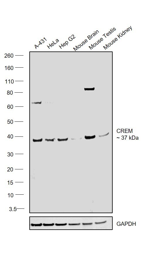
- Western blot was performed using Anti-CREM Polyclonal Antibody(Product # PA5-29927) and a 37 kDa band corresponding to CREM was observed across the cell lines and tissues tested. Whole Cell Extract (1% SDS), (30 µg lysate) of A-431 (Lane 1), HeLa (Lane 2), Hep G2 (Lane 3), Mouse Brain (Lane 4), Mouse Testis (Lane 5) and Mouse Kidney (Lane 6) were electrophoresed using NuPAGE™ 4-12% Bis-Tris Protein Gel (Product # NP0321BOX). Resolved proteins were then transferred onto a Nitrocellulose membrane (Product # LC2001) by iBlot® 2 Dry Blotting System (Product # IB21001). The blot was probed with the primary antibody (1:1000 dilution) and detected by chemiluminescence with Goat anti-Rabbit IgG (H+L) Superclonal™ Recombinant Secondary Antibody, HRP (Product # A27036, 1:4000 dilution) using the iBright FL 1000 (Product # A32752). Chemiluminescent detection was performed using Novex® ECL Chemiluminescent Substrate Reagent Kit (Product # WP20005).
Supportive validation
- Submitted by
- Invitrogen Antibodies (provider)
- Main image
- Experimental details
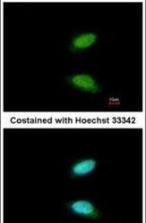
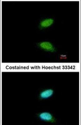
- Immunofluorescent analysis of CREM in paraformaldehyde-fixed HeLa cells using a CREM polyclonal antibody (Product # PA5-29927) at a 1:200 dilution.
Supportive validation
- Submitted by
- Invitrogen Antibodies (provider)
- Main image
- Experimental details
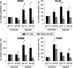
- Fig. 2 Change of protein levels of PER3, GLS2, CREM and TXNIP in AT1 prostate and Walker-256 mammary carcinoma cells after 24 h under acidotic (pH 6.6) and/or hypoxic (pO 2 = 1.5 mmHg) conditions. n = 4-12; (*) p < 0.05, (**) p < 0.01 vs. control; ( # ) p < 0.05, ( ## ) p < 0.01 AT1 vs. Walker-256 cells

- Submitted by
- Invitrogen Antibodies (provider)
- Main image
- Experimental details
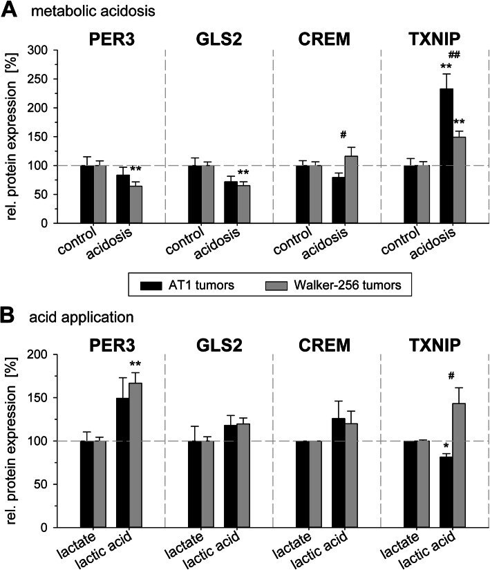
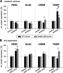
- Fig. 4 Protein levels of PER3, GLS2, CREM and TXNIP in AT1 prostate and Walker-256 mammary carcinoma tumors in vivo 24 h after ( a ) inducing metabolic acidosis by forcing glycolytic metabolism or ( b ) intratumoral injection of lactic acid. n = 4-18; (*) p < 0.05, (**) p < 0.01 vs. control; ( # ) p < 0.05, ( ## ) p < 0.01 AT1 vs. Walker-256 cells