 Explore
 Validate
 Learn
Learn Western blot
Antibody data
- Antibody Data
- Antigen structure
- References [2]
- Comments [0]
- Validations
- Western blot [4]
- Immunocytochemistry [1]
- Immunohistochemistry [1]
- Other assay [2]
Submit
Validation data
Reference
Comment
Report error
- Product number
- PA5-21387 - Provider product page

- Provider
- Invitrogen Antibodies
- Product name
- ENO1 Polyclonal Antibody
- Antibody type
- Polyclonal
- Antigen
- Recombinant protein fragment
- Description
- Recommended positive controls: 293T, A431, H1299, HeLa, HepG2, Molt-4, Raji, mouse brain. Predicted reactivity: Mouse (94%), Rat (93%), Xenopus laevis (90%), Chicken (93%), Rhesus Monkey (100%), Chimpanzee (100%), Bovine (93%), Guinea pig (95%). Store product as a concentrated solution. Centrifuge briefly prior to opening the vial.
- Reactivity
- Human, Mouse
- Host
- Rabbit
- Isotype
- IgG
- Vial size
- 100 µL
- Concentration
- 1 mg/mL
- Storage
- Store at 4°C short term. For long term storage, store at -20°C, avoiding freeze/thaw cycles.
Submitted references Quantitative Proteomic Analysis of Differentially Expressed Protein Profiles Involved in Pancreatic Ductal Adenocarcinoma.
Proteomic analysis of the effects of aged garlic extract and its FruArg component on lipopolysaccharide-induced neuroinflammatory response in microglial cells.
Kuo KK, Kuo CJ, Chiu CY, Liang SS, Huang CH, Chi SW, Tsai KB, Chen CY, Hsi E, Cheng KH, Chiou SH
Pancreas 2016 Jan;45(1):71-83
Pancreas 2016 Jan;45(1):71-83
Proteomic analysis of the effects of aged garlic extract and its FruArg component on lipopolysaccharide-induced neuroinflammatory response in microglial cells.
Zhou H, Qu Z, Mossine VV, Nknolise DL, Li J, Chen Z, Cheng J, Greenlief CM, Mawhinney TP, Brown PN, Fritsche KL, Hannink M, Lubahn DB, Sun GY, Gu Z
PloS one 2014;9(11):e113531
PloS one 2014;9(11):e113531
No comments: Submit comment
Supportive validation
- Submitted by
- Invitrogen Antibodies (provider)
- Main image
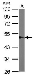
- Experimental details
- Western Blot using ENO1 Polyclonal Antibody (Product # PA5-21387). Sample (50 µg of whole cell lysate). Lane A: Mouse brain. 10% SDS PAGE. ENO1 Polyclonal Antibody (Product # PA5-21387) diluted at 1:1,000. The HRP-conjugated anti-rabbit IgG antibody was used to detect the primary antibody.

- Submitted by
- Invitrogen Antibodies (provider)
- Main image

- Experimental details
- Western Blot using ENO1 Polyclonal Antibody (Product # PA5-21387). Sample (30 µg of whole cell lysate). Lane A: A431. Lane B: H1299. 10% SDS PAGE. ENO1 Polyclonal Antibody (Product # PA5-21387) diluted at 1:3,000. The HRP-conjugated anti-rabbit IgG antibody was used to detect the primary antibody.

- Submitted by
- Invitrogen Antibodies (provider)
- Main image

- Experimental details
- Western Blot using ENO1 Polyclonal Antibody (Product # PA5-21387). Various whole cell extracts (30 µg) were separated by 10% SDS-PAGE, and the membrane was blotted with ENO1 Polyclonal Antibody (Product # PA5-21387) diluted at 1:1,000. The HRP-conjugated anti-rabbit IgG antibody was used to detect the primary antibody.

- Submitted by
- Invitrogen Antibodies (provider)
- Main image

- Experimental details
- Western blot analysis was performed on whole cell extracts (30 µg lysate) of C2C12 (Lane 1), Hep G2 (Lane 2), NIH/3T3 (Lane 3) and MCF7 (Lane 4). The blot was probed with Anti-ENO1 Polyclonal Antibody (Product # PA5-21387, 1 µg/mL) and detected by chemiluminescence using Goat anti-Rabbit IgG (H+L) Superclonal™ Secondary Antibody, HRP conjugate (Product # A27036, 0.25 µg/mL, 1:4000 dilution). A 50 kDa band corresponding to ENO1 was observed across the cell lines tested.

Supportive validation
- Submitted by
- Invitrogen Antibodies (provider)
- Main image

- Experimental details
- Immunocytochemistry-Immunofluorescence analysis of ENO1 was performed in HeLa cells fixed in 4% paraformaldehyde at RT for 15 min. Green: ENO1 Polyclonal Antibody (Product # PA5-21387) diluted at 1:200. Red: alpha Tubulin, a cytoskeleton marker. Blue: Hoechst 33342 staining.

Supportive validation
- Submitted by
- Invitrogen Antibodies (provider)
- Main image

- Experimental details
- Immunohistochemical analysis of paraffin-embedded human gastric tissue, using ENO1 (Product # PA5-21387) antibody at 1:100 dilution. Antigen Retrieval: EDTA based buffer, pH 8.0, 15 min.

Supportive validation
- Submitted by
- Invitrogen Antibodies (provider)
- Main image

- Experimental details
- Figure 5 Validation of expression profiling of proteins by Western blotting. Four of the identified proteins, PRDX1 ( A ), GLRX3 ( B ), ENO1 ( C ), and CASP1 ( D ), responding to AGE and/or FruArg treatment in LPS-stimulated BV-2 cells were validated using Western blotting. Protein intensities were quantified by Image J software, normalized to beta-actin, and expressed as percentage of untreated controls. Data are means +- SEM (n>=5); #, P

- Submitted by
- Invitrogen Antibodies (provider)
- Main image

- Experimental details
- FIGURE 5 The differentially expressed proteins with upregulated patterns at the indicated time points in the normal and tumor-bearing mice were identified by comparative proteome analysis using nano-LC-MS/MS that corresponds to the results analyzed by quantitative real-time PCR and Western blot. All values were normalized to the appropriate control groups, which were set to 1.0. T indicates tumor-bearing mice; N, normal control mice; the indicated time points of the 1 to 4 groups are 10, 5, 3.5, and 2.5 weeks. A, The 5 representative upregulated proteins were dramatically accompanied with PDAC progression in tumor-bearing mice. B, The 5 representative mRNA expression levels examined by quantitative real-time PCR increased concomitantly with PDAC progression. C, The 3 representative upregulated proteins were confirmed and examined by Western blot. D, The 3 representative upregulated proteins were confirmed and examined by IHC staining.
