Antibody data
- Antibody Data
- Antigen structure
- References [2]
- Comments [0]
- Validations
- Western blot [1]
- ELISA [1]
- Immunocytochemistry [2]
- Other assay [1]
Submit
Validation data
Reference
Comment
Report error
- Product number
- MA1-169 - Provider product page

- Provider
- Invitrogen Antibodies
- Product name
- TGF beta-1 Monoclonal Antibody (B11-4C3)
- Antibody type
- Monoclonal
- Antigen
- Other
- Description
- MA1-169 has been successfully used as a coating antibody in a sandwich ELISA with Product # MA1-116 used as a detection antibody.
- Reactivity
- Human
- Host
- Mouse
- Isotype
- IgG
- Antibody clone number
- B11-4C3
- Vial size
- 200 µg
- Concentration
- 1 mg/mL
- Storage
- -20°C
Submitted references Histological changes of cervical tumours following Zanthoxylum acanthopodium DC treatment, and its impact on cytokine expression.
LncRNA MCTP1-AS1 Regulates EMT Process in Endometrial Cancer by Targeting the miR-650/SMAD7 Axis.
Simanullang RH, Situmorang PC, Herlina M, Noradina, Silalahi B, Manurung SS
Saudi journal of biological sciences 2022 Apr;29(4):2706-2718
Saudi journal of biological sciences 2022 Apr;29(4):2706-2718
LncRNA MCTP1-AS1 Regulates EMT Process in Endometrial Cancer by Targeting the miR-650/SMAD7 Axis.
Gao Q, Huang Q, Li F, Luo F
OncoTargets and therapy 2021;14:751-761
OncoTargets and therapy 2021;14:751-761
No comments: Submit comment
Supportive validation
- Submitted by
- Invitrogen Antibodies (provider)
- Main image

- Experimental details
- Western blot analysis of human TGF beta-1 was performed by loading 10 µL of human serum from patients with multiple sclerosis (MS), leukemia, or asthma (left panel) and 2 µg and 1 µg of recombinant human TGF beta-1 protein (right panel) per well on a 4-20% Tris-HCl polyacrylamide gel. Proteins were transferred to a PVDF membrane and blocked with StartingBlock (TBS) Blocking Buffer (Product # 37542) for at least 1 hour. The membrane was probed with a TGF beta-1 monoclonal antibody (Product # MA1-169) at a concentration of 5 µg/mL overnight at 4C on a rocking platform, washed in TBS-0.1%Tween-20, and probed with a goat anti-mouse IgG-HRP secondary antibody (Product # 31430) at a dilution of 1:10,000 for at least 1 hour. Chemiluminescent detection was performed using SuperSignal West Dura (Product # 34075).
Supportive validation
- Submitted by
- Invitrogen Antibodies (provider)
- Main image

- Experimental details
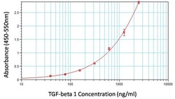
- Sandwich ELISA analysis of an anti-human TGF beta-1 monoclonal antibody (Product # MA1-169) was performed by loading 100 µL per well of antibody (Product # MA1-169) at a concentration of 2 µg/mL overnight at room temperature. The plate was washed 3 times with ELISA Wash Buffer (Product # N503), and 100 µL of recombinant human TGF beta-1 was added to wells in duplicate at 2.5, 1.2, 0.625, 0.312, 0.156, 0.078, 0.039 and 0 ng/mL concentrations and the samples were incubated for 2 hours at room temperature. The plate was washed, then incubated with 100 µL per well of a biotinylated TGF beta-1 monoclonal antibody (Product # MA1-116, biotinylated using EZ-Link Sulfo-NHS-LC-Biotinylation Kit (Product # 21435) at a concentration of 0.125 µg/mL for 1 hour at room temperature, followed by 100 µL per well of Streptavidin-HRP (Product # N504) at a dilution of 1:40,000 for 30 minutes at room temperature. Detection was performed by adding 100 µL of 1-Step Ultra TMB substrate (Product # 34028) per well and incubating for 20 minutes at room temperature in the dark. The plate was then stopped with 100 µL per well of 0.16M sulfuric acid. Absorbances were read on a spectrophotometer at 450-550 nm.
Supportive validation
- Submitted by
- Invitrogen Antibodies (provider)
- Main image

- Experimental details
- Immunofluorescence analysis of TGF beta1 was performed using THP-1 and THP-1 cells differentiated and polarized to M2 macrophages. The cells were fixed with 4% paraformaldehyde for 10 minutes, permeabilized with 0.1% Triton™ X-100 for 10 minutes, and blocked with 2% BSA for 45 minutes at room temperature. The cells were labeled with TGF beta-1 Monoclonal Antibody (B11-4C3) (Product # MA1-169) at 1:100 in 0.1% BSA, incubated at 4 degree celsius overnight and then labeled with Donkey anti-Mouse IgG (H+L) Highly Cross-Adsorbed Secondary Antibody, Alexa Fluor Plus 488 (Product # A32766), (1:2500), for 45 minutes at room temperature (Panel a: Green). Nuclei (Panel b: Blue) were stained with ProLong™ Diamond Antifade Mountant with DAPI (Product # P36962). F-actin (Panel c: Red) was stained with Rhodamine Phalloidin (Product # R415, 1:300). Panel d represents the merged image showing cytoplasmic localization of TGF beta1 in M2 macrophages compared to THP-1 control cells (Panel e). Panel f represents control M2 macrophage cells with no primary antibody to assess background. The images were captured at 60X magnification.
- Submitted by
- Invitrogen Antibodies (provider)
- Main image

- Experimental details
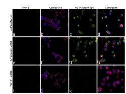
- Knockdown of Transforming growth factor beta-1 proprotein was achieved by transfecting THP-1 cells with TGF beta1 specific siRNA (Silencer® select Product # s14056, s14054). Immunofluorescence analysis was performed on untransfected THP1 cells (panel a,b), untransfected M2 macrophage (panel c, d), non-specific scrambled siRNA transfected THP-1 (panels e,f), non-specific scrambled siRNA transfected M2 macrophage (panels g,h), THP-1 transfected with TGF beta1 specific siRNA (panel i,j), and THP-1 transfected with TGF beta1 specific siRNA and differentiated to M2 macrophage (panels k,l) (Green). Cells were fixed, permeabilized, and labelled with TGF beta-1 Monoclonal Antibody (B11-4C3) (Product # MA1-169, 1:100) followed by Donkey anti-Mouse IgG (H+L) Highly Cross-Adsorbed Secondary Antibody, Alexa Fluor Plus 488 (Product # A32766), (1:2500 dilution). Nuclei (blue) were stained using ProLong™ Diamond Antifade Mountant with DAPI (Product # P36962), and Rhodamine Phalloidin (Product # R415, 1:300) was used for cytoskeletal F-actin (Red) staining. Reduction of specific signal was observed upon siRNA mediated knockdown (panel k,l) confirming specificity of the antibody to TGF beta1. The Images were captured at 60X magnification.
Supportive validation
- Submitted by
- Invitrogen Antibodies (provider)
- Main image

- Experimental details
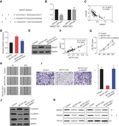
- Figure 5 MCTP1-AS1 regulated SMAD7 expression via miR-650 in EC cells. (A) The binding scheme of miR-650 and SMAD7. ( B ) miR-650 interacts with SMAD7 by directly targeting verified by luciferase reporter assay. Data are mean +- SD of triplicate experiment (***p
 Explore
 Validate
 Learn
Learn Western blot