 Explore
 Validate
 Learn
Learn Western blot
Antibody data
- Antibody Data
- Antigen structure
- References [2]
- Comments [0]
- Validations
- Western blot [4]
- Immunohistochemistry [2]
- Other assay [2]
Submit
Validation data
Reference
Comment
Report error
- Product number
- MA5-23891 - Provider product page

- Provider
- Invitrogen Antibodies
- Product name
- Cathepsin L Monoclonal Antibody (204101)
- Antibody type
- Monoclonal
- Antigen
- Recombinant full-length protein
- Description
- In direct ELISAs and Western blots, no cross-reactivity with recombinant human Cathepsin B, C, L2, O, S, or X/Z/P is observed. In Western blots, both the pro and active forms of recombinant human and mouse Cathspsin L are recognized.
- Antibody clone number
- 204101
- Concentration
- 0.5 mg/mL
Submitted references Cardiac and Renal SARS-CoV-2 Viral Entry Protein Regulation by Androgens and Diet: Implications for Polycystic Ovary Syndrome and COVID-19.
SARS-CoV-2 Viral Entry Proteins in Hyperandrogenemic Female Mice: Implications for Women with PCOS and COVID-19.
Rezq S, Huffman AM, Basnet J, Yanes Cardozo LL, Romero DG
International journal of molecular sciences 2021 Sep 9;22(18)
International journal of molecular sciences 2021 Sep 9;22(18)
SARS-CoV-2 Viral Entry Proteins in Hyperandrogenemic Female Mice: Implications for Women with PCOS and COVID-19.
Huffman AM, Rezq S, Basnet J, Yanes Cardozo LL, Romero DG
International journal of molecular sciences 2021 Apr 25;22(9)
International journal of molecular sciences 2021 Apr 25;22(9)
No comments: Submit comment
Supportive validation
- Submitted by
- Invitrogen Antibodies (provider)
- Main image

- Experimental details
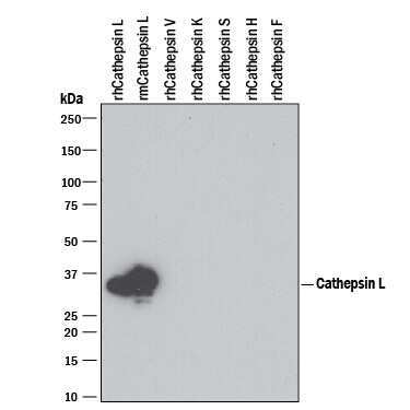
- Western blot analysis of Cathepsin L in 100 ng recombinant Human Cathepsin L, recombinant Mouse Cathepsin L , recombinant Human Cathepsin V, recombinant Human Cathepsin K, recombinant Human Cathepsin S, recombinant Human Cathepsin H, and recombinant Human Cathepsin F. Samples were incubated in Cathepsin L monoclonal antibody (Product # MA5-23891) using a dilution of 1 µg/mL followed by a HRP-conjugated Anti-Rat IgG secondary antibody. A specific band was detected for Cathepsin L at approximately 35 kDa (as indicated). This experiment was conducted under reducing conditions.
- Submitted by
- Invitrogen Antibodies (provider)
- Main image

- Experimental details
- Western blot analysis of Cathepsin L in 100 ng recombinant Human Cathepsin L, recombinant Mouse Cathepsin L , recombinant Human Cathepsin V, recombinant Human Cathepsin K, recombinant Human Cathepsin S, recombinant Human Cathepsin H, and recombinant Human Cathepsin F. Samples were incubated in Cathepsin L monoclonal antibody (Product # MA5-23891) using a dilution of 1 µg/mL followed by a HRP-conjugated Anti-Rat IgG secondary antibody. A specific band was detected for Cathepsin L at approximately 35 kDa (as indicated). This experiment was conducted under reducing conditions.

- Submitted by
- Invitrogen Antibodies (provider)
- Main image

- Experimental details
- Knockdown of Cathepsin L was achieved by transfecting A549 with Cathepsin L specific siRNAs (Silencer® select Product # S223364, S3754). Western blot analysis (Fig. a) was performed using Whole cell extracts from the Cathepsin L knockdown cells (lane 3), non-targeting scrambled siRNA transfected cells (lane 2) and untransfected cells (lane 1). The blot was probed with Cathepsin L Monoclonal Antibody (204101) (Product # MA5-23891, 1 ug/ml ) and F(ab2-Rabbit anti-Rat IgG (H+L Secondary Antibody, HRP (Product # PA1-29927, 1:4000 dilution). Densitometric analysis of this western blot is shown in histogram (Fig. b). Decrease in signal upon siRNA mediated knock down confirms that antibody is specific to Cathepsin L.

- Submitted by
- Invitrogen Antibodies (provider)
- Main image
- Experimental details
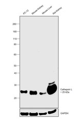
- Western blot was performed using Anti-Cathepsin L Monoclonal Antibody (204101)(Product # MA5-23891) and a 25kDa band corresponding to mature form of Cathepsin L was observed across cell line and tissue extracts tested. Whole cell extract (30 µg lysate) of PC-12 (Lane 1) and tissue extracts of Mouse Kidney (Lane 2), Mouse Liver (Lane 3) and Rat Kidney (Lane 4) were electrophoresed using NuPAGE™ 4-12% Bis-Tris Protein Gel (Product # NP0322BOX). Resolved proteins were then transferred onto a Nitrocellulose membrane (Product # IB23001) by iBlot® 2 Dry Blotting System (Product # IB21001). The blot was probed with the primary antibody (1 ug/ml) and detected by chemiluminescence with F(ab2-Rabbit anti-Rat IgG (H+L Secondary Antibody, HRP (Product # PA1-29927,1:4000 dilution) using the iBright FL 1000 (Product # A32752). Chemiluminescent detection was performed using SuperSignal™ West Dura Extended Duration Substrate (Product # 34076).

Supportive validation
- Submitted by
- Invitrogen Antibodies (provider)
- Main image

- Experimental details
- Immunohistochemical analysis of Cathepsin L in immersion fixed paraffin-embedded sections of human kidney. Samples were incubated in Cathepsin L monoclonal antibody (Product # MA5-23891) using a dilution of 5 µg/mL overnight at 4 °C. Tissue was stained using the Anti-Rat HRP-DAB Cell & Tissue Staining Kit (brown) and counterstained with hematoxylin (blue). Specific staining was localized to cytoplasm in tubular epithelial cells.

- Submitted by
- Invitrogen Antibodies (provider)
- Main image

- Experimental details
- Immunohistochemical analysis of Cathepsin L in immersion fixed frozen sections of mouse kidney. Samples were incubated in Cathepsin L monoclonal antibody (Product # MA5-23891) using a dilution of 8 µg/mL overnight at 4 °C. Tissue was stained using the Anti-Rat HRP-DAB Cell & Tissue Staining Kit (brown) and counterstained with hematoxylin (blue). Specific staining was localized to cytoplasm in tubular epithelial cells.

Supportive validation
- Submitted by
- Invitrogen Antibodies (provider)
- Main image
- Experimental details
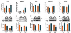
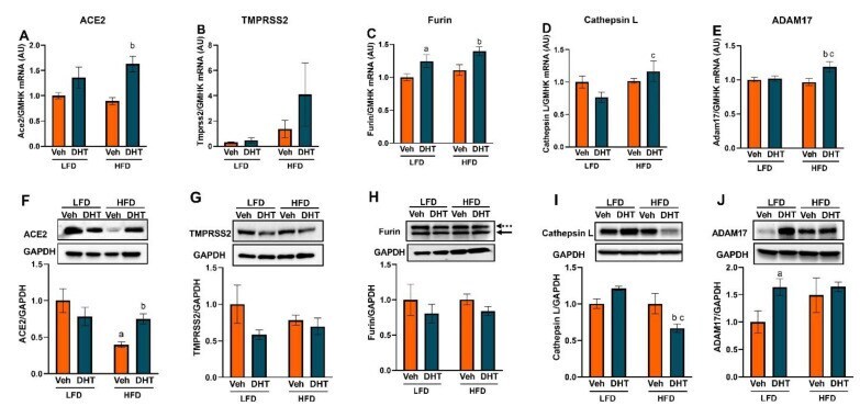
- Figure 3 Effect of DHT and diet on cardiac SARS-CoV-2 viral entry proteins expression. Animals were treated with dihydrotestosterone (DHT) or vehicle (Veh) and maintained in low (LFD) or high (HFD) fat diet for 90 days. Ace2 ( A ), Tmprss2 ( B ), furin ( C ), cathepsin L ( D ), and ADAM17 ( E ) mRNA was quantified by RT-qPCR and standardized to the geometric mean of four housekeeping genes (HGMK) ( N = 6/group). ACE2 ( F ), TMPRSS2 ( G ), furin ( H ), cathepsin L ( I ), and ADAM17 ( J ) protein was quantified by Western-blot and normalized to GAPDH ( N = 4/group). Total furin ( H ) was quantified as the sum of profurin (dashed arrow) and cleaved soluble furin (solid arrow). Data are expressed as mean +- SEM. Data were analyzed by two-way ANOVA followed by Fisher's LSD test. a p < 0.05 vs. LFD-Veh; b p < 0.05 vs. HFD-Veh; c p < 0.05 vs. LFD-DHT.
- Submitted by
- Invitrogen Antibodies (provider)
- Main image
- Experimental details
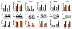
- Figure 4 Effect of DHT and diet on renal SARS-CoV-2 viral entry proteins expression. Animals were treated with dihydrotestosterone (DHT) or vehicle (Veh) and maintained in low (LFD) or high (HFD) fat diet for 90 days. Ace2 ( A ), Tmprss2 ( B ), Tmprss4 ( C ), furin ( D ), cathepsin L ( E ), and ADAM17 ( F ) mRNA was quantified by RT-qPCR and standardized to the geometric mean of four housekeeping genes (HGMK) ( N = 6-8/group). ACE2 ( G ), TMPRSS2 ( H ), TMPRSS4 ( I ), furin ( J ), cathepsin L ( K ), and ADAM17 ( L ) protein was quantified by Western-blot and normalized to GAPDH ( N = 4/group). Total furin ( J ) was quantified as the sum of profurin (dashed arrow) and cleaved soluble furin (solid arrow). Data are expressed as mean +- SEM. Data were analyzed by two-way ANOVA followed by Fisher's LSD test. a p < 0.05 vs. LFD-Veh; b p < 0.05 vs. LFD-Veh; c p < 0.05 vs. LFD-DHT.
