Explore
 Validate
 Learn
Learn Western blot
 Immunoprecipitation
Antibody data
- Antibody Data
- Antigen structure
- References [2]
- Comments [0]
- Validations
- Western blot [2]
- Blocking/Neutralizing [1]
Submit
Validation data
Reference
Comment
Report error
- Product number
- MAB1408 - Provider product page

- Provider
- R&D Systems
- Product name
- Human Cystatin B Antibody
- Antibody type
- Monoclonal
- Description
- Protein A or G purified from hybridoma culture supernatant. Detects human Cystatin B in Western blots and direct ELISAs. In Western blots, less than 5% cross-reactivity with recombinant human (rh) Cystatins A and S, recombinant mouse Cystatin B, rhFetuin A, and rhHPRG (Histidine-Proline-Rich Glycoprotein) is observed and approximately 5% to 10% cross-reactivity with rhCystatins C, D, E/M, SA, SN, and rhFetuin B is observed.
- Reactivity
- Human
- Host
- Mouse
- Conjugate
- Unconjugated
- Antigen sequence
P04080- Isotype
- IgG
- Antibody clone number
- 225228
- Vial size
- 500 ug
- Storage
- Use a manual defrost freezer and avoid repeated freeze-thaw cycles. 12 months from date of receipt, -20 to -70 °C as supplied. 1 month, 2 to 8 °C under sterile conditions after reconstitution. 6 months, -20 to -70 °C under sterile conditions after reconstitution.
Submitted references Dysregulation of macrophage-secreted cathepsin B contributes to HIV-1-linked neuronal apoptosis.
Cystatin B as a tissue and urinary biomarker of bladder cancer recurrence and disease progression.
Rodriguez-Franco EJ, Cantres-Rosario YM, Plaud-Valentin M, Romeu R, Rodríguez Y, Skolasky R, Meléndez V, Cadilla CL, Melendez LM
PloS one 2012;7(5):e36571
PloS one 2012;7(5):e36571
Cystatin B as a tissue and urinary biomarker of bladder cancer recurrence and disease progression.
Feldman AS, Banyard J, Wu CL, McDougal WS, Zetter BR
Clinical cancer research : an official journal of the American Association for Cancer Research 2009 Feb 1;15(3):1024-31
Clinical cancer research : an official journal of the American Association for Cancer Research 2009 Feb 1;15(3):1024-31
No comments: Submit comment
Supportive validation
- Submitted by
- R&D Systems (provider)
- Main image
- Experimental details
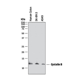
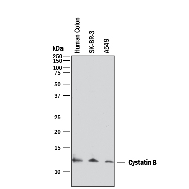
- Detection of Human Cystatin B by Western Blot. Western blot shows lysates of human colon tissue, SK-BR-3 human breast cancer cell line, and A549 human lung carcinoma cell line. PVDF membrane was probed with 2 µg/mL of Mouse Anti-Human Cystatin B Monoclonal Antibody (Catalog # MAB1408) followed by HRP-conjugated Anti-Mouse IgG Secondary Antibody (Catalog # HAF018). A specific band was detected for Cystatin B at approximately 12 kDa (as indicated). This experiment was conducted under reducing conditions and using Immunoblot Buffer Group 1.
- Submitted by
- R&D Systems (provider)
- Main image
- Experimental details
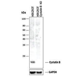
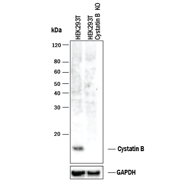
- Western Blot Shows Human Cystatin B Specificity by Using Knockout Cell Line. Western blot shows lysates of HEK293T human embryonic kidney parental cell line and Cystatin B knockout HEK293T cell line (KO). PVDF membrane was probed with 2 µg/mL of Mouse Anti-Human Cystatin B Monoclonal Antibody (Catalog # MAB1408) followed by HRP-conjugated Anti-Mouse IgG Secondary Antibody (Catalog # HAF018). A specific band was detected for Cystatin B at approximately 12 kDa (as indicated) in the parental HEK293T cell line, but is not detectable in knockout HEK293T cell line. GAPDH (Catalog # MAB5718) is shown as a loading control. This experiment was conducted under reducing conditions and using Immunoblot Buffer Group 1.
Supportive validation
- Submitted by
- R&D Systems (provider)
- Main image

- Experimental details
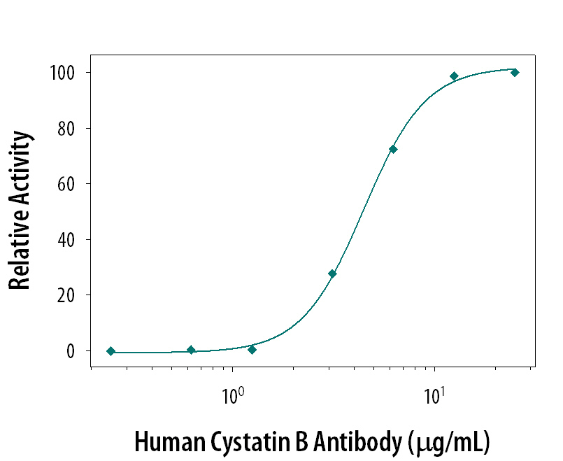
- Neutralization of Cystatin B Activity by Human Cystatin B Antibody. Papain (0.1 µg/mL) activity is measured in the presence of Recombinant Human Cystatin B (0.53 µg/mL, Catalog # 1408-PI) that has been preincubated with increasing concentrations of Human Cystatin B Monoclonal Antibody (Catalog # MAB1408). The ND50 is typically 5.4 µg/mL.