 Explore
 Validate
 Learn
LearnGTX25476
antibody from GeneTex
Targeting: FGFR2
BEK, CD332, CEK3, CFD1, ECT1, JWS, K-SAM, KGFR, TK14, TK25
 Western blot
Antibody data
- Antibody Data
- Antigen structure
- References [4]
- Comments [0]
- Validations
- Western blot [1]
- Immunocytochemistry [1]
- Immunohistochemistry [3]
- Flow cytometry [1]
Submit
Validation data
Reference
Comment
Report error
- Product number
- GTX25476 - Provider product page

- Provider
- GeneTex
- Proper citation
- GeneTex Cat#GTX25476, RRID:AB_380552
- Product name
- FGFR2 antibody
- Antibody type
- Polyclonal
- Reactivity
- Human, Mouse
- Host
- Rabbit
Submitted references Runx2 is required for the proliferation of osteoblast progenitors and induces proliferation by regulating Fgfr2 and Fgfr3.
Transcriptomics and Transposon Mutagenesis Identify Multiple Mechanisms of Resistance to the FGFR Inhibitor AZD4547.
The FGFR/MEK/ERK/brachyury pathway is critical for chordoma cell growth and survival.
Targeting FGFR4 inhibits hepatocellular carcinoma in preclinical mouse models.
Kawane T, Qin X, Jiang Q, Miyazaki T, Komori H, Yoshida CA, Matsuura-Kawata VKDS, Sakane C, Matsuo Y, Nagai K, Maeno T, Date Y, Nishimura R, Komori T
Scientific reports 2018 Sep 10;8(1):13551
Scientific reports 2018 Sep 10;8(1):13551
Transcriptomics and Transposon Mutagenesis Identify Multiple Mechanisms of Resistance to the FGFR Inhibitor AZD4547.
Kas SM, de Ruiter JR, Schipper K, Schut E, Bombardelli L, Wientjens E, Drenth AP, de Korte-Grimmerink R, Mahakena S, Phillips C, Smith PD, Klarenbeek S, van de Wetering K, Berns A, Wessels LFA, Jonkers J
Cancer research 2018 Oct 1;78(19):5668-5679
Cancer research 2018 Oct 1;78(19):5668-5679
The FGFR/MEK/ERK/brachyury pathway is critical for chordoma cell growth and survival.
Hu Y, Mintz A, Shah SR, Quinones-Hinojosa A, Hsu W
Carcinogenesis 2014 Jul;35(7):1491-9
Carcinogenesis 2014 Jul;35(7):1491-9
Targeting FGFR4 inhibits hepatocellular carcinoma in preclinical mouse models.
French DM, Lin BC, Wang M, Adams C, Shek T, Hötzel K, Bolon B, Ferrando R, Blackmore C, Schroeder K, Rodriguez LA, Hristopoulos M, Venook R, Ashkenazi A, Desnoyers LR
PloS one 2012;7(5):e36713
PloS one 2012;7(5):e36713
No comments: Submit comment
Supportive validation
- Submitted by
- GeneTex (provider)
- Main image
- Experimental details
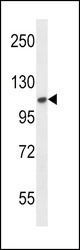
- FGFR2 Antibody (N-term) (GTX25476) western blot analysis in mouse NIH-3T3 cell line lysates (35μg/lane). This demonstrates the FGFR2 antibody detected the FGFR2 protein (arrow).

Supportive validation
- Submitted by
- GeneTex (provider)
- Main image
- Experimental details
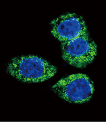
- Confocal immunofluorescent analysis of FGFR2 Antibody (N-term) (GTX25476) with Hela cell followed by Alexa Fluor 488-conjμgated goat anti-rabbit lgG (green).DAPI was used to stain the cell nuclear (blue).

Supportive validation
- Submitted by
- GeneTex (provider)
- Main image
- Experimental details
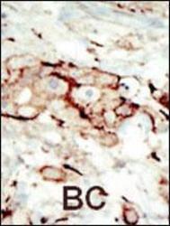
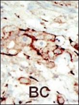
- Formalin-fixed and paraffin-embedded human cancer tissue reacted with the primary FGFR2 Antibody (N-term) (GTX25476) , which was peroxidase-conjμgated to the secondary antibody, followed by AEC staining. This data demonstrates the use of this antibody for immunohistochemistry.BC = breast carcinoma
- Submitted by
- GeneTex (provider)
- Main image
- Experimental details
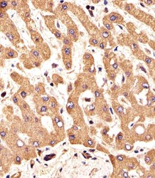
- Immunohistochemical analysis of paraffin-embedded H. liver section using FGFR2 Antibody (N-term)(GTX25476). GTX25476 was diluted at 1:25 dilution. A undiluted biotinylated goat polyvalent antibody was used as the secondary, followed by DAB staining.

- Submitted by
- GeneTex (provider)
- Main image
- Experimental details
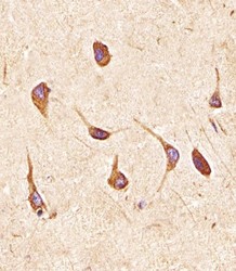
- Immunohistochemical analysis of paraffin-embedded H. brain section using FGFR2 Antibody (N-term)(GTX25476). GTX25476 was diluted at 1:25 dilution. A undiluted biotinylated goat polyvalent antibody was used as the secondary, followed by DAB staining.

Supportive validation
- Submitted by
- GeneTex (provider)
- Main image
- Experimental details
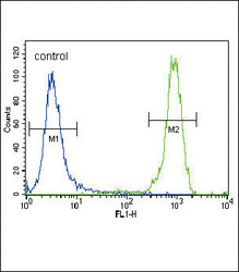
- FGFR2 Antibody (N-term) (GTX25476) flow cytometric analysis of NCI-H460 cells (right histogram) compared to a negative control cell (left histogram). FITC-conjμgated goat-anti-rabbit secondary antibodies were used for the analysis.
