 Explore
 Validate
 Learn
Learn Western blot
 Immunocytochemistry
Antibody data
- Antibody Data
- Antigen structure
- References [2]
- Comments [0]
- Validations
- Western blot [2]
- Immunocytochemistry [1]
- Immunohistochemistry [8]
Submit
Validation data
Reference
Comment
Report error
- Product number
- HPA022845 - Provider product page

- Provider
- Atlas Antibodies
- Proper citation
- Atlas Antibodies Cat#HPA022845, RRID:AB_1854399
- Product name
- Anti-NEFM
- Antibody type
- Polyclonal
- Reactivity
- Human
- Host
- Rabbit
- Conjugate
- Unconjugated
- Antigen sequence
KEEEEKEVKEAPKEEKVEKKEEKPKDVPEKKKAES
PVKEEAVAEVVTITKSVKVHLEKETKEEGKPLQQE
KEKEKAGGEGGSEEEGSDKGAKG- Isotype
- IgG
- Vial size
- 100 µl
- Storage
- Store at +4°C for short term storage. Long time storage is recommended at -20°C.
Submitted references Plasma profiling reveals three proteins associated to amyotrophic lateral sclerosis.
Affinity Proteomics Reveals Elevated Muscle Proteins in Plasma of Children with Cerebral Malaria
Häggmark A, Mikus M, Mohsenchian A, Hong MG, Forsström B, Gajewska B, Barańczyk-Kuźma A, Uhlén M, Schwenk JM, Kuźma-Kozakiewicz M, Nilsson P
Annals of clinical and translational neurology 2014 Aug;1(8):544-53
Annals of clinical and translational neurology 2014 Aug;1(8):544-53
Affinity Proteomics Reveals Elevated Muscle Proteins in Plasma of Children with Cerebral Malaria
Bachmann J, Burté F, Pramana S, Conte I, Brown B, Orimadegun A, Ajetunmobi W, Afolabi N, Akinkunmi F, Omokhodion S, Akinbami F, Shokunbi W, Kampf C, Pawitan Y, Uhlén M, Sodeinde O, Schwenk J, Wahlgren M, Fernandez-Reyes D, Nilsson P, Kim K
PLoS Pathogens 2014 April;10(4)
PLoS Pathogens 2014 April;10(4)
No comments: Submit comment
Enhanced validation
Enhanced validation
- Submitted by
- Atlas Antibodies (provider)
- Enhanced method
- Orthogonal validation
- Main image

- Experimental details
- Western blot analysis in human cell line HEK 293 and human cell line A-431.

Enhanced validation
- Submitted by
- Atlas Antibodies (provider)
- Enhanced method
- Independent antibody validation
- Main image

- Experimental details
- Western blot analysis using Anti-NEFM antibody HPA022845 (A) shows similar pattern to independent antibody HPA023138 (B).

Supportive validation
- Submitted by
- Atlas Antibodies (provider)
- Main image

- Experimental details
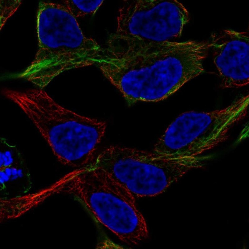
- Immunofluorescent staining of human cell line HEK 293 shows localization to intermediate filaments.
- Sample type
- HUMAN
Enhanced validation
Supportive validation
- Submitted by
- Atlas Antibodies (provider)
- Enhanced method
- Orthogonal validation
- Main image

- Experimental details
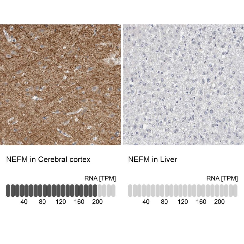
- Immunohistochemistry analysis in human cerebral cortex and liver tissues using HPA022845 antibody. Corresponding NEFM RNA-seq data are presented for the same tissues.
- Sample type
- HUMAN
Supportive validation
- Submitted by
- Atlas Antibodies (provider)
- Main image

- Experimental details
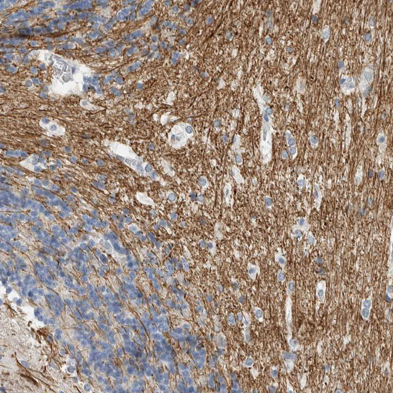
- Immunohistochemical staining of human cerebellum shows strong cytoplasmic positivity.

- Submitted by
- Atlas Antibodies (provider)
- Main image

- Experimental details
- Immunohistochemical staining of human cerebral cortex shows high expression.
- Sample type
- HUMAN

- Submitted by
- Atlas Antibodies (provider)
- Main image

- Experimental details
- Immunohistochemical staining of human pancreas shows low expression as expected.
- Sample type
- HUMAN

- Submitted by
- Atlas Antibodies (provider)
- Main image

- Experimental details
- Immunohistochemical staining of human cerebellum shows strong cytoplasmic positivity in white matter.
- Sample type
- HUMAN
- Submitted by
- Atlas Antibodies (provider)
- Main image

- Experimental details
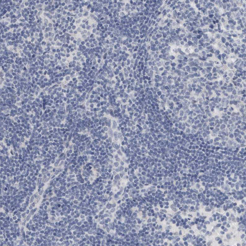
- Immunohistochemical staining of human lymph node shows no positivity in non-germinal center cells as expected.
- Sample type
- HUMAN
- Submitted by
- Atlas Antibodies (provider)
- Main image

- Experimental details
- Immunohistochemical staining of human liver shows no positivity in hepatocytes as expected.
- Sample type
- HUMAN

- Submitted by
- Atlas Antibodies (provider)
- Main image

- Experimental details
- Immunohistochemical staining of human cerebral cortex shows strong cytoplasmic positivity in neurons.
- Sample type
- HUMAN
