Explore
 Validate
 Learn
Learn Western blot
 Immunocytochemistry
Antibody data
- Antibody Data
- Antigen structure
- References [2]
- Comments [0]
- Validations
- Western blot [5]
- Immunocytochemistry [1]
- Immunohistochemistry [1]
Submit
Validation data
Reference
Comment
Report error
- Product number
- GTX103323 - Provider product page

- Provider
- GeneTex
- Proper citation
- GeneTex Cat#GTX103323, RRID:AB_1951292
- Product name
- T-Plastin antibody
- Antibody type
- Polyclonal
- Reactivity
- Human, Mouse
- Host
- Rabbit
Submitted references Effect of genetic background on the phenotype of the Smn2B/- mouse model of spinal muscular atrophy.
PLS3 mutations in X-linked osteoporosis with fractures.
Eshraghi M, McFall E, Gibeault S, Kothary R
Human molecular genetics 2016 Oct 15;25(20):4494-4506
Human molecular genetics 2016 Oct 15;25(20):4494-4506
PLS3 mutations in X-linked osteoporosis with fractures.
van Dijk FS, Zillikens MC, Micha D, Riessland M, Marcelis CL, de Die-Smulders CE, Milbradt J, Franken AA, Harsevoort AJ, Lichtenbelt KD, Pruijs HE, Rubio-Gozalbo ME, Zwertbroek R, Moutaouakil Y, Egthuijsen J, Hammerschmidt M, Bijman R, Semeins CM, Bakker AD, Everts V, Klein-Nulend J, Campos-Obando N, Hofman A, te Meerman GJ, Verkerk AJ, Uitterlinden AG, Maugeri A, Sistermans EA, Waisfisz Q, Meijers-Heijboer H, Wirth B, Simon ME, Pals G
The New England journal of medicine 2013 Oct 17;369(16):1529-36
The New England journal of medicine 2013 Oct 17;369(16):1529-36
No comments: Submit comment
Enhanced validation
Supportive validation
- Submitted by
- GeneTex (provider)
- Enhanced method
- Genetic validation
- Main image

- Experimental details
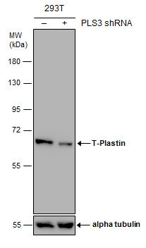
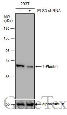
- Non-transfected (¡V) and transfected (+) 293T whole cell extracts (30 ?g) were separated by 7.5% SDS-PAGE, and the membrane was blotted with T-Plastin antibody (GTX103323) diluted at 1:4000. The HRP-conjugated anti-rabbit IgG antibody (GTX213110-01) was used to detect the primary antibody.
Supportive validation
- Submitted by
- GeneTex (provider)
- Main image

- Experimental details
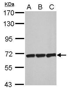
- Sample (30 ?g of whole cell lysate) A: NIH-3T3 B: JC C: BCL-1 7.5% SDS PAGE GTX103323 diluted at 1:1000 The HRP-conjugated anti-rabbit IgG antibody (GTX213110-01) was used to detect the primary antibody.
- Submitted by
- GeneTex (provider)
- Main image
- Experimental details
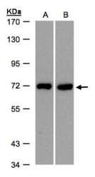
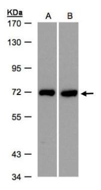
- Sample(30 ug whole cell lysate)A:HeLa S3(GTX14654)B:Raji(GTX27908)7.5% SDS PAGEGTX103323 diluted at 1:500
- Validation comment
- WB
- Submitted by
- GeneTex (provider)
- Main image

- Experimental details
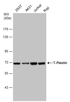
- T-Plastin antibody detects T-Plastin protein by western blot analysis. Various whole cell extracts (30 ?g) were separated by 7.5% SDS-PAGE, and the membrane was blotted with T-Plastin antibody (GTX103323) diluted at a dilution of 1:1000. The HRP-conjugated anti-rabbit IgG antibody (GTX213110-01) was used to detect the primary antibody.
- Submitted by
- GeneTex (provider)
- Main image

- Experimental details
- Non-transfected (¡V) and transfected (+) 293T whole cell extracts (30 ?g) were separated by 7.5% SDS-PAGE, and the membrane was blotted with T-Plastin antibody (GTX103323) diluted at 1:4000. The HRP-conjugated anti-rabbit IgG antibody (GTX213110-01) was used to detect the primary antibody.
Supportive validation
- Submitted by
- GeneTex (provider)
- Main image
- Experimental details
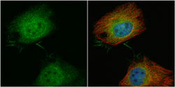
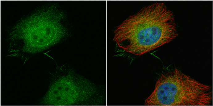
- T-Plastin antibody detects T-Plastin protein at cytoplasm and nucleus by immunofluorescent analysis.Sample: HeLa cells were fixed in 4% paraformaldehyde at RT for 15 min.Green: T-Plastin protein stained by T-Plastin antibody (GTX103323) diluted at 1:100.Red: alpha Tubulin, a cytoskeleton marker, stained by alpha Tubulin antibody [GT114] (GTX628802) diluted at 1:500.Blue: Hoechst 33342 staining.
Supportive validation
- Submitted by
- GeneTex (provider)
- Main image

- Experimental details
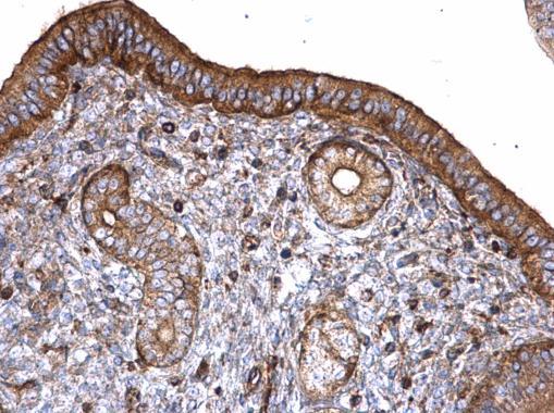
- T-Plastin antibody detects T-Plastin protein at cytoplasm on mouse cervix by immunohistochemical analysis. Sample: Paraffin-embedded mouse cervix. T-Plastin antibody (GTX103323) diluted at 1:500.