 Explore
 Validate
 Learn
Learn Western blot
 Immunocytochemistry
Antibody data
- Antibody Data
- Antigen structure
- References [1]
- Comments [0]
- Validations
- Western blot [3]
- Immunocytochemistry [1]
- Immunohistochemistry [2]
Submit
Validation data
Reference
Comment
Report error
- Product number
- GTX632482 - Provider product page

- Provider
- GeneTex
- Product name
- T-Plastin antibody [GT3310]
- Antibody type
- Monoclonal
- Reactivity
- Human, Mouse, Rat
- Host
- Mouse
Submitted references Angiotensin II regulates phosphorylation of actin-associated proteins in human podocytes.
Schenk LK, Möller-Kerutt A, Klosowski R, Wolters D, Schaffner-Reckinger E, Weide T, Pavenstädt H, Vollenbröker B
FASEB journal : official publication of the Federation of American Societies for Experimental Biology 2017 Nov;31(11):5019-5035
FASEB journal : official publication of the Federation of American Societies for Experimental Biology 2017 Nov;31(11):5019-5035
No comments: Submit comment
Enhanced validation
Supportive validation
- Submitted by
- GeneTex (provider)
- Enhanced method
- Genetic validation
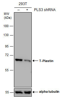
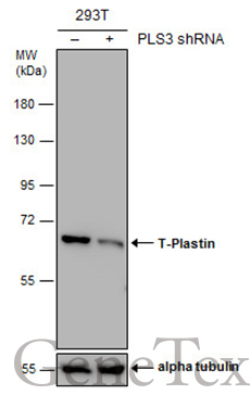
- Main image

- Experimental details
- Non-transfected (¡V) and transfected (+) 293T whole cell extracts (30 ?g) were separated by 7.5% SDS-PAGE, and the membrane was blotted with T-Plastin antibody [GT3310] (GTX632482) diluted at 1:1000.
Supportive validation
- Submitted by
- GeneTex (provider)
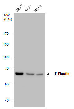
- Main image

- Experimental details
- T-Plastin antibody detects T-Plastin protein by western blot analysis. Various whole cell extracts (30 £gg) were separated by 7.5% SDS-PAGE, and the membrane was blotted with T-Plastin antibody (GTX632482) diluted at a dilution of 1:1000.
- Submitted by
- GeneTex (provider)
- Main image
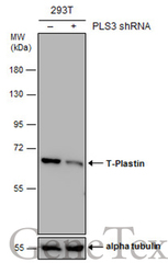
- Experimental details
- Non-transfected (¡V) and transfected (+) 293T whole cell extracts (30 ?g) were separated by 7.5% SDS-PAGE, and the membrane was blotted with T-Plastin antibody [GT3310] (GTX632482) diluted at 1:1000.
Supportive validation
- Submitted by
- GeneTex (provider)
- Main image
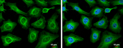
- Experimental details
- T-Plastin antibody [GT3310] detects T-Plastin protein at cytoplasm by immunofluorescent analysis.Sample: HeLa cells were fixed in 4% paraformaldehyde at RT for 15 min.Green: T-Plastin protein stained by T-Plastin antibody [GT3310] (GTX632482) diluted at 1:200.Blue: Hoechst 33342 staining.Scale bar = 10 £gm.

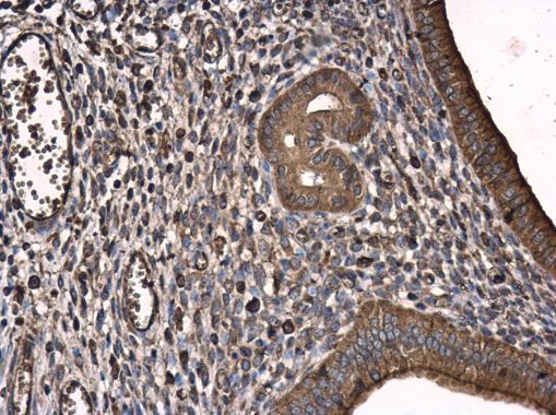
Supportive validation
- Submitted by
- GeneTex (provider)
- Main image
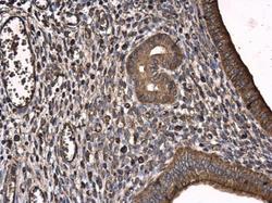
- Experimental details
- T-Plastin antibody [GT3310] detects T-Plastin protein at cytoplasm in mouse cervix by immunohistochemical analysis. Sample: Paraffin-embedded mouse cervix. T-Plastin antibody [GT3310] (GTX632482) diluted at 1:250.
- Submitted by
- GeneTex (provider)
- Main image
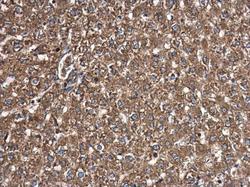
- Experimental details
- T-Plastin antibody [GT3310] detects T-Plastin protein at cytoplasm in rat liver by immunohistochemical analysis. Sample: Paraffin-embedded rat liver. T-Plastin antibody [GT3310] (GTX632482) diluted at 1:250.
