Explore
 Validate
 Learn
Learn Western blot
Antibody data
- Antibody Data
- Antigen structure
- References [5]
- Comments [0]
- Validations
- Western blot [3]
- Immunocytochemistry [1]
- Immunohistochemistry [1]
- Other assay [1]
Submit
Validation data
Reference
Comment
Report error
- Product number
- PA5-27883 - Provider product page

- Provider
- Invitrogen Antibodies
- Product name
- PLS3 Polyclonal Antibody
- Antibody type
- Polyclonal
- Antigen
- Recombinant protein fragment
- Reactivity
- Human, Mouse
- Host
- Rabbit
- Isotype
- IgG
- Vial size
- 100 µL
- Concentration
- 0.23 mg/mL
- Storage
- Store at 4°C short term. For long term storage, store at -20°C, avoiding freeze/thaw cycles.
Submitted references Cul3 regulates cytoskeleton protein homeostasis and cell migration during a critical window of brain development.
T-Plastin reinforces membrane protrusions to bridge matrix gaps during cell migration.
Evaluation of the role of an antioxidant gene in NSC-34 motor neuron-like cells as a model of a motor neuron disease.
The Actin Binding Protein Plastin-3 Is Involved in the Pathogenesis of Acute Myeloid Leukemia.
Circulating tumour cell-derived plastin3 is a novel marker for predicting long-term prognosis in patients with breast cancer.
Morandell J, Schwarz LA, Basilico B, Tasciyan S, Dimchev G, Nicolas A, Sommer C, Kreuzinger C, Dotter CP, Knaus LS, Dobler Z, Cacci E, Schur FKM, Danzl JG, Novarino G
Nature communications 2021 May 24;12(1):3058
Nature communications 2021 May 24;12(1):3058
T-Plastin reinforces membrane protrusions to bridge matrix gaps during cell migration.
Garbett D, Bisaria A, Yang C, McCarthy DG, Hayer A, Moerner WE, Svitkina TM, Meyer T
Nature communications 2020 Sep 23;11(1):4818
Nature communications 2020 Sep 23;11(1):4818
Evaluation of the role of an antioxidant gene in NSC-34 motor neuron-like cells as a model of a motor neuron disease.
Alrafiah AR
Folia morphologica 2019;78(1):1-9
Folia morphologica 2019;78(1):1-9
The Actin Binding Protein Plastin-3 Is Involved in the Pathogenesis of Acute Myeloid Leukemia.
Velthaus A, Cornils K, Hennigs JK, Grüb S, Stamm H, Wicklein D, Bokemeyer C, Heuser M, Windhorst S, Fiedler W, Wellbrock J
Cancers 2019 Oct 26;11(11)
Cancers 2019 Oct 26;11(11)
Circulating tumour cell-derived plastin3 is a novel marker for predicting long-term prognosis in patients with breast cancer.
Ueo H, Sugimachi K, Gorges TM, Bartkowiak K, Yokobori T, Müller V, Shinden Y, Ueda M, Ueo H, Mori M, Kuwano H, Maehara Y, Ohno S, Pantel K, Mimori K
British journal of cancer 2015 Apr 28;112(9):1519-26
British journal of cancer 2015 Apr 28;112(9):1519-26
No comments: Submit comment
Supportive validation
- Submitted by
- Invitrogen Antibodies (provider)
- Main image
- Experimental details
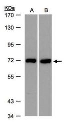
- Western blot analysis of T-Plastin using 30 µg of A) HeLa S3 and B) Raji lysate. Samples were loaded onto a 7.5% SDS-PAGE gel and probed with a T-Plastin polyclonal antibody (Product # PA5-27883) at a dilution of 1:500.
- Submitted by
- Invitrogen Antibodies (provider)
- Main image
- Experimental details
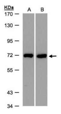
- Western Blot using PLS3 Polyclonal Antibody (Product # PA5-27883). Sample (30 µg of whole cell lysate). Lane A: NIH-3T3. Lane B: JC. Lane C: BCL-1. 7.5% SDS PAGE. PLS3 Polyclonal Antibody (Product # PA5-27883) diluted at 1:1,000. The HRP-conjugated anti-rabbit IgG antibody was used to detect the primary antibody.

- Submitted by
- Invitrogen Antibodies (provider)
- Main image
- Experimental details
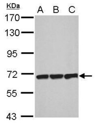
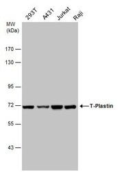
- PLS3 Polyclonal Antibody detects T-Plastin protein by western blot analysis. Various whole cell extracts (30 µg) were separated by 7.5% SDS-PAGE, and the membrane was blotted with PLS3 Polyclonal Antibody (Product # PA5-27883) diluted at a dilution of 1:1,000. The HRP-conjugated anti-rabbit IgG antibody was used to detect the primary antibody.

Supportive validation
- Submitted by
- Invitrogen Antibodies (provider)
- Main image
- Experimental details
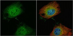
- Immunocytochemistry-Immunofluorescence analysis of PLS3 was performed in HeLa cells fixed in 4% paraformaldehyde at RT for 15 min. Green: PLS3 Polyclonal Antibody (Product # PA5-27883) diluted at 1:100. Red: alpha Tubulin, a cytoskeleton marker. Blue: Hoechst 33342 staining.

Supportive validation
- Submitted by
- Invitrogen Antibodies (provider)
- Main image

- Experimental details
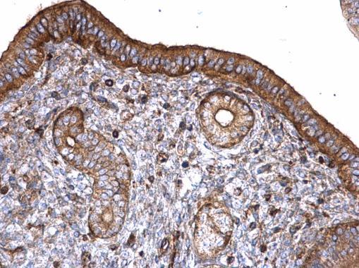
- PLS3 Polyclonal Antibody detects T-Plastin protein at cytoplasm on mouse cervix by immunohistochemical analysis. Sample: Paraffin-embedded mouse cervix. PLS3 Polyclonal Antibody (Product # PA5-27883) diluted at 1:500. Antigen Retrieval: EDTA based buffer, pH 8.0, 15 min.
Supportive validation
- Submitted by
- Invitrogen Antibodies (provider)
- Main image
- Experimental details
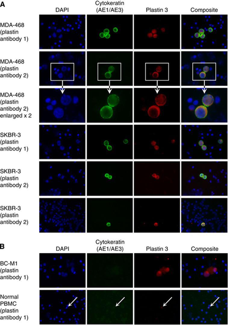
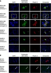
- Figure 2 Comparison of PLS3 expression in breast cancer cell lines with PLS3 expression in peripheral blood mononuclear cells of healthy control individuals by immunocytochemical double staining. Cells of the assigned cell lines were spiked in the blood samples. Identification of the tumour cells was supported by the cytokeratin-specific antibody AE1/AE3. Nuclei were stained with DAPI, and the composite image is an overlay of the DAPI, cytokeratin, and PLS3 images. Two different PLS3 antibodies were applied for the analysis. ( A ) For MDA-MB-468 (MDA-468), an enlarged view of stained cells is shown. ( B ) BC-M1 analysis for PLS3 and blood samples without cell spiking. PLS3 signals in PBMC were labelled with arrows.