 Explore
 Validate
 Learn
Learn Immunocytochemistry
Antibody data
- Antibody Data
- Antigen structure
- References [4]
- Comments [0]
- Validations
- Immunocytochemistry [2]
- Immunohistochemistry [5]
Submit
Validation data
Reference
Comment
Report error
- Product number
- HPA024223 - Provider product page

- Provider
- Atlas Antibodies
- Proper citation
- Atlas Antibodies Cat#HPA024223, RRID:AB_1854248
- Product name
- Anti-MYO10
- Antibody type
- Polyclonal
- Reactivity
- Human
- Host
- Rabbit
- Conjugate
- Unconjugated
- Antigen sequence
QRMKEQQELSLTEASLQKLQERRDQELRRLEEEAC
RAAQEFLESLNFDEIDECVRNIERSLSVGSEFSSE
LAESACEEKPNFNFSQPYPEEEVDEGFEADDDAFK
DSPNPSEHGHSDQRTSGIRTSDDSSEEDPYMNDTV
VPTSPSA- Isotype
- IgG
- Vial size
- 100 µl
- Storage
- Store at +4°C for short term storage. Long time storage is recommended at -20°C.
Submitted references DPP6 regulation of dendritic morphogenesis impacts hippocampal synaptic development.
Myosin X and its motorless isoform differentially modulate dendritic spine development by regulating trafficking and retention of vasodilator-stimulated phosphoprotein.
Myosin-X functions in polarized epithelial cells.
Headless Myo10 is a negative regulator of full-length Myo10 and inhibits axon outgrowth in cortical neurons.
Lin L, Sun W, Throesch B, Kung F, Decoster JT, Berner CJ, Cheney RE, Rudy B, Hoffman DA
Nature communications 2013;4:2270
Nature communications 2013;4:2270
Myosin X and its motorless isoform differentially modulate dendritic spine development by regulating trafficking and retention of vasodilator-stimulated phosphoprotein.
Lin WH, Hurley JT, Raines AN, Cheney RE, Webb DJ
Journal of cell science 2013 Oct 15;126(Pt 20):4756-68
Journal of cell science 2013 Oct 15;126(Pt 20):4756-68
Myosin-X functions in polarized epithelial cells.
Liu KC, Jacobs DT, Dunn BD, Fanning AS, Cheney RE
Molecular biology of the cell 2012 May;23(9):1675-87
Molecular biology of the cell 2012 May;23(9):1675-87
Headless Myo10 is a negative regulator of full-length Myo10 and inhibits axon outgrowth in cortical neurons.
Raines AN, Nagdas S, Kerber ML, Cheney RE
The Journal of biological chemistry 2012 Jul 20;287(30):24873-83
The Journal of biological chemistry 2012 Jul 20;287(30):24873-83
No comments: Submit comment
Supportive validation
- Submitted by
- Atlas Antibodies (provider)
- Main image
- Experimental details
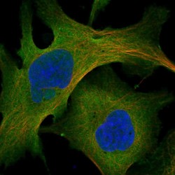
- Immunofluorescent staining of human cell line U-2 OS shows localization to plasma membrane & cytosol.
- Sample type
- HUMAN

- Submitted by
- Atlas Antibodies (provider)
- Main image
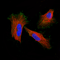
- Experimental details
- Immunofluorescent staining of human cell line HeLa shows localization to plasma membrane and the tips of filopodia. Antibody staining is shown in green.
- Sample type
- HUMAN

Supportive validation
- Submitted by
- Atlas Antibodies (provider)
- Main image

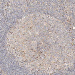
- Experimental details
- Immunohistochemical staining of human skeletal muscle shows moderate cytopalsmic positivity in myocytes.

- Submitted by
- Atlas Antibodies (provider)
- Main image
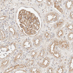
- Experimental details
- Immunohistochemical staining of human kidney shows strong membranous positivity in cells in glomeruli.
- Sample type
- HUMAN

- Submitted by
- Atlas Antibodies (provider)
- Main image
- Experimental details
- Immunohistochemical staining of human lymphoid tissues shows weak cytoplasmic positivity in germinal center cells.
- Sample type
- HUMAN

- Submitted by
- Atlas Antibodies (provider)
- Main image
- Experimental details
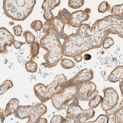
- Immunohistochemical staining of human placenta shows moderate cytoplasmic positivity in trophoblastic cells.
- Sample type
- HUMAN

- Submitted by
- Atlas Antibodies (provider)
- Main image
- Experimental details
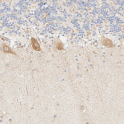
- Immunohistochemical staining of human cerebellum shows moderate cytoplasmic positivity in Purkinje cells.
- Sample type
- HUMAN
