Explore
 Validate
 Learn
Learn17-1719-41
antibody from Invitrogen Antibodies
Targeting: L1CAM
CD171, HSAS, HSAS1, MASA, MIC5, S10, SPG1
 Flow cytometry
Antibody data
- Antibody Data
- Antigen structure
- References [1]
- Comments [0]
- Validations
- Flow cytometry [2]
- Other assay [1]
Submit
Validation data
Reference
Comment
Report error
- Product number
- 17-1719-41 - Provider product page

- Provider
- Invitrogen Antibodies
- Product name
- CD171 Monoclonal Antibody (eBio5G3 (5G3)), APC, eBioscience™
- Antibody type
- Monoclonal
- Antigen
- Other
- Description
- The monoclonal antibody eBio5G3 recognizes CD171 also known as neural cell adhesion molecule L1. CD171 is a member of the Ig superfamily containing 6 extracellular Ig domains and five fibronectin type III-like repeats. CD171 has been shown to function as a cell adhesion molecule mediating homotypic and heterotypic cell-cell interactions in neuronal myelination, neurite outgrowth and regeneration. Expression of CD171 has been found on monocytes and mature monocytic-derived and follicular DCs, a minor subset of lymphocytes in addition to that found on neuronal tissue and some tumor cells lines. Expression of CD171 on tumors is thought to contribute to tumor progression. Epitope of eBio5G3 is in amino-terminal Ig-like domain. This eBio5G3 (5G3) antibody has been pre-titrated and tested by flow cytometric analysis of the tumor cell line Panc-1. This can be used at 5 µL (0.06 µg) per test. A test is defined as the amount (µg) of antibody that will stain a cell sample in a final volume of 100 µL. Cell number should be determined empirically but can range from 105 to 108 cells/test. Filtration: 0.2 µm post-manufacturing filtered.
- Reactivity
- Human
- Host
- Mouse
- Isotype
- IgG
- Antibody clone number
- eBio5G3 (5G3)
- Vial size
- 25 Tests
- Concentration
- 5 µL/Test
- Storage
- 4° C, store in dark, DO NOT FREEZE!
Submitted references Plasma Levels of Neuron- and Astrocyte-Derived Exosomal Amyloid Beta1-42, Amyloid Beta1-40, and Phosphorylated Tau Levels in Schizophrenia Patients and Non-psychiatric Comparison Subjects: Relationships With Cognitive Functioning and Psychopathology.
Lee EE, Winston-Gray C, Barlow JW, Rissman RA, Jeste DV
Frontiers in psychiatry 2020;11:532624
Frontiers in psychiatry 2020;11:532624
No comments: Submit comment
Supportive validation
- Submitted by
- Invitrogen Antibodies (provider)
- Main image

- Experimental details
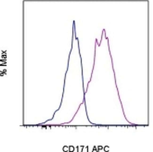
- Staining of Panc-1 cells with Mouse IgG2a K Isotype Control APC (Product # 17-4724) (blue histogram) or Anti-Human CD171 APC (purple histogram). Total viable cells were used for analysis.

- Submitted by
- Invitrogen Antibodies (provider)
- Main image

- Experimental details
- Staining of Panc-1 cells with Mouse IgG2a K Isotype Control APC (Product # 17-4724) (blue histogram) or Anti-Human CD171 APC (purple histogram). Total viable cells were used for analysis.
Supportive validation
- Submitted by
- Invitrogen Antibodies (provider)
- Main image
- Experimental details
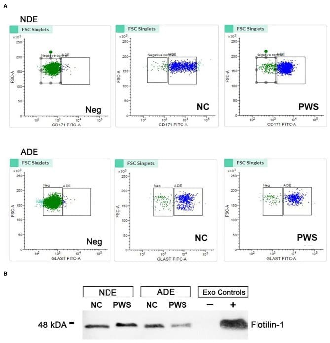
- Figure 1 Characterization of neuronal (NDE) and astrocyte derived exosomes (ADE)s. (A) FACS analysis of plasma NDEs and ADEs following the formation of bead-antibody-exosome (BAE)-FITC complexes. Streptavidin magnetic beads were incubated with exosomes isolated from non-psychiatric comparison subjects (NC) and people with schizophrenia (PWS) ( n = 60) and enriched against biotinylated anti-human CD171 biotin (L1CAM, NDE) or anti-GLAST antibody (ADE). BAE complexes are stained with FITC prior to FACS. (B) Plasma NDE and ADE preparations from NC and PWS patients were probed with exosome marker, Flotilin-1 (1:1000). Non-exosome fraction (supernant resulting from 1 h spin at 1500G) served as the negative control while total exosomes (diluted in 1x PBS prior to neuronal and astrocyte enrichment) served as the positive control. The resultant Western Blot demonstrated that NDEs are Flotillin and Neun positive and GFAP negative. However, we were not able to get a signal for the ADEs. Due to multiple freeze-thaw cycles, the integrity of the samples used in the current study had diminished significantly.