Explore
 Validate
 Learn
Learn Western blot
 Immunocytochemistry
Antibody data
- Antibody Data
- Antigen structure
- References [7]
- Comments [0]
- Validations
- Western blot [2]
Submit
Validation data
Reference
Comment
Report error
- Product number
- AF3184 - Provider product page

- Provider
- R&D Systems
- Product name
- Human/Mouse HMGA2 Antibody
- Antibody type
- Polyclonal
- Description
- Immunogen affinity purified. Detects human and mouse HMGA2 in direct ELISAs and Western blots. In direct ELISAs, approximately 10% cross-reactivity with recombinant human HMGB1 is observed.
- Reactivity
- Human, Mouse
- Host
- Goat
- Conjugate
- Unconjugated
- Antigen sequence
P52926- Isotype
- IgG
- Vial size
- 100 ug
- Concentration
- LYOPH
- Storage
- Use a manual defrost freezer and avoid repeated freeze-thaw cycles. 12 months from date of receipt, -20 to -70 °C as supplied. 1 month, 2 to 8 °C under sterile conditions after reconstitution. 6 months, -20 to -70 °C under sterile conditions after reconstitution.
Submitted references Multicolor quantitative confocal imaging cytometry.
Generation and characterisation of a canine EGFP-HMGA2 prostate cancer in vitro model.
HMGA1 and HMGA2 expression and comparative analyses of HMGA2, Lin28 and let-7 miRNAs in oral squamous cell carcinoma.
MiRNA let-7g regulates skeletal myoblast motility via Pinch-2.
Downregulation of HMGA2 by the pan-deacetylase inhibitor panobinostat is dependent on hsa-let-7b expression in liver cancer cell lines.
Subsets of very low risk Wilms tumor show distinctive gene expression, histologic, and clinical features.
Clinical significance of high mobility group A2 in human gastric cancer and its relationship to let-7 microRNA family.
Coutu DL, Kokkaliaris KD, Kunz L, Schroeder T
Nature methods 2018 Jan;15(1):39-46
Nature methods 2018 Jan;15(1):39-46
Generation and characterisation of a canine EGFP-HMGA2 prostate cancer in vitro model.
Willenbrock S, Wagner S, Reimann-Berg N, Moulay M, Hewicker-Trautwein M, Nolte I, Murua Escobar H
PloS one 2014;9(6):e98788
PloS one 2014;9(6):e98788
HMGA1 and HMGA2 expression and comparative analyses of HMGA2, Lin28 and let-7 miRNAs in oral squamous cell carcinoma.
Sterenczak KA, Eckardt A, Kampmann A, Willenbrock S, Eberle N, Länger F, Kleinschmidt S, Hewicker-Trautwein M, Kreipe H, Nolte I, Murua Escobar H, Gellrich NC
BMC cancer 2014 Sep 23;14:694
BMC cancer 2014 Sep 23;14:694
MiRNA let-7g regulates skeletal myoblast motility via Pinch-2.
Boudoukha S, Rivera Vargas T, Dang I, Kropp J, Cuvellier S, Gautreau A, Polesskaya A
FEBS letters 2014 May 2;588(9):1623-9
FEBS letters 2014 May 2;588(9):1623-9
Downregulation of HMGA2 by the pan-deacetylase inhibitor panobinostat is dependent on hsa-let-7b expression in liver cancer cell lines.
Di Fazio P, Montalbano R, Neureiter D, Alinger B, Schmidt A, Merkel AL, Quint K, Ocker M
Experimental cell research 2012 Sep 10;318(15):1832-43
Experimental cell research 2012 Sep 10;318(15):1832-43
Subsets of very low risk Wilms tumor show distinctive gene expression, histologic, and clinical features.
Sredni ST, Gadd S, Huang CC, Breslow N, Grundy P, Green DM, Dome JS, Shamberger RC, Beckwith JB, Perlman EJ, Renal Tumor Committee of the Children's Oncology Group.
Clinical cancer research : an official journal of the American Association for Cancer Research 2009 Nov 15;15(22):6800-9
Clinical cancer research : an official journal of the American Association for Cancer Research 2009 Nov 15;15(22):6800-9
Clinical significance of high mobility group A2 in human gastric cancer and its relationship to let-7 microRNA family.
Motoyama K, Inoue H, Nakamura Y, Uetake H, Sugihara K, Mori M
Clinical cancer research : an official journal of the American Association for Cancer Research 2008 Apr 15;14(8):2334-40
Clinical cancer research : an official journal of the American Association for Cancer Research 2008 Apr 15;14(8):2334-40
No comments: Submit comment
Supportive validation
- Submitted by
- R&D Systems (provider)
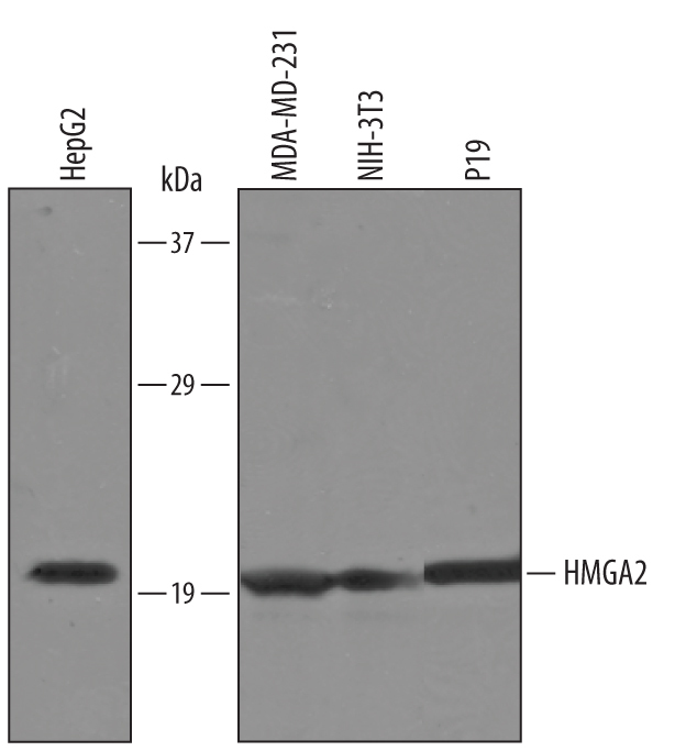
- Main image

- Experimental details
- Detection of Human and Mouse HMGA2 by Western Blot. Western blot shows lysates of HepG2 human hepatocellular carcinoma cell line, MDA-MD-231 human breast cancer cell line, NIH-3T3 mouse embryonic fibroblast cell line, and P19 mouse embryonal carcinoma cell line. PVDF Membrane was probed with 1 µg/mL of Goat Anti-Human/Mouse HMGA2 Antigen Affinity-purified Polyclonal Antibody (Catalog # AF3184) followed by HRP-conjugated Anti-Goat IgG Secondary Antibody (Catalog # HAF019). A specific band was detected for HMGA2 at approximately 21 kDa (as indicated). This experiment was conducted under reducing conditions and using Immunoblot Buffer Group 8.
- Submitted by
- R&D Systems (provider)
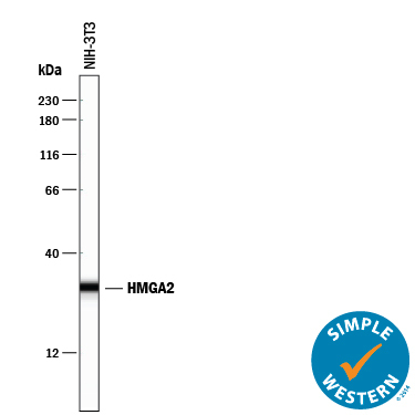
- Main image

- Experimental details
- Detection of Mouse HMGA2 by Simple WesternTM. Simple Western lane view shows lysates of NIH-3T3 mouse embryonic fibroblast cell line, loaded at 0.2 mg/mL. A specific band was detected for HMGA2 at approximately 30 kDa (as indicated) using 10 µg/mL of Goat Anti-Human/Mouse HMGA2 Antigen Affinity-purified Polyclonal Antibody (Catalog # AF3184) followed by 1:50 dilution of HRP-conjugated Anti-Goat IgG Secondary Antibody (Catalog # HAF109). This experiment was conducted under reducing conditions and using the 12-230 kDa separation system.