Antibody data
- Antibody Data
- Antigen structure
- References [6]
- Comments [0]
- Validations
- Immunohistochemistry [1]
Submit
Validation data
Reference
Comment
Report error
- Product number
- MAB923-100 - Provider product page

- Provider
- R&D Systems
- Product name
- Human Angiopoietin-1 Antibody
- Antibody type
- Monoclonal
- Description
- Protein A or G purified from hybridoma culture supernatant. Detects human Angiopoietin-1 in direct ELISAs and Western blots. In direct ELISAs and Western blots, no cross-reactivity with recombinant human (rh) Angiopoietin-2, recombinant mouse Angiopoietin-like 3, rhAngiopoietin-4, or rhAngiopoietin-like 7 is observed.
- Reactivity
- Human
- Host
- Mouse
- Conjugate
- Unconjugated
- Antigen sequence
Q15389- Isotype
- IgG
- Antibody clone number
- 171718
- Vial size
- 100 ug
- Storage
- Use a manual defrost freezer and avoid repeated freeze-thaw cycles. 12 months from date of receipt, -20 to -70 °C as supplied. 1 month, 2 to 8 °C under sterile conditions after reconstitution. 6 months, -20 to -70 °C under sterile conditions after reconstitution.
Submitted references Pulmonary pericytes regulate lung morphogenesis.
Fibulin-5 Regulates Angiopoietin-1/Tie-2 Receptor Signaling in Endothelial Cells.
Early systemic microvascular damage in pigs with atherogenic diabetes mellitus coincides with renal angiopoietin dysbalance.
High bone marrow angiopoietin-1 expression is an independent poor prognostic factor for survival in patients with myelodysplastic syndromes.
Regulation of angiopoietin expression by bacterial lipopolysaccharide.
Angiopoietin 1 directly induces destruction of the rheumatoid joint by cooperative, but independent, signaling via ERK/MAPK and phosphatidylinositol 3-kinase/Akt.
Kato K, Diéguez-Hurtado R, Park DY, Hong SP, Kato-Azuma S, Adams S, Stehling M, Trappmann B, Wrana JL, Koh GY, Adams RH
Nature communications 2018 Jun 22;9(1):2448
Nature communications 2018 Jun 22;9(1):2448
Fibulin-5 Regulates Angiopoietin-1/Tie-2 Receptor Signaling in Endothelial Cells.
Chan W, Ismail H, Mayaki D, Sanchez V, Tiedemann K, Davis EC, Hussain SN
PloS one 2016;11(6):e0156994
PloS one 2016;11(6):e0156994
Early systemic microvascular damage in pigs with atherogenic diabetes mellitus coincides with renal angiopoietin dysbalance.
Khairoun M, van den Heuvel M, van den Berg BM, Sorop O, de Boer R, van Ditzhuijzen NS, Bajema IM, Baelde HJ, Zandbergen M, Duncker DJ, Rabelink TJ, Reinders ME, van der Giessen WJ, Rotmans JI
PloS one 2015;10(4):e0121555
PloS one 2015;10(4):e0121555
High bone marrow angiopoietin-1 expression is an independent poor prognostic factor for survival in patients with myelodysplastic syndromes.
Cheng CL, Hou HA, Jhuang JY, Lin CW, Chen CY, Tang JL, Chou WC, Tseng MH, Yao M, Huang SY, Ko BS, Hsu SC, Wu SJ, Tsay W, Chen YC, Tien HF
British journal of cancer 2011 Sep 27;105(7):975-82
British journal of cancer 2011 Sep 27;105(7):975-82
Regulation of angiopoietin expression by bacterial lipopolysaccharide.
Mofarrahi M, Nouh T, Qureshi S, Guillot L, Mayaki D, Hussain SN
American journal of physiology. Lung cellular and molecular physiology 2008 May;294(5):L955-63
American journal of physiology. Lung cellular and molecular physiology 2008 May;294(5):L955-63
Angiopoietin 1 directly induces destruction of the rheumatoid joint by cooperative, but independent, signaling via ERK/MAPK and phosphatidylinositol 3-kinase/Akt.
Hashiramoto A, Sakai C, Yoshida K, Tsumiyama K, Miura Y, Shiozawa K, Nose M, Komai K, Shiozawa S
Arthritis and rheumatism 2007 Jul;56(7):2170-9
Arthritis and rheumatism 2007 Jul;56(7):2170-9
No comments: Submit comment
Supportive validation
- Submitted by
- R&D Systems (provider)
- Main image

- Experimental details
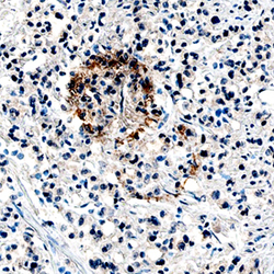
- Angiopoietin-1 in Human Prostate Cancer Tissue. Angiopoietin-1 was detected in immersion fixed paraffin-embedded sections of human prostate cancer tissue using Mouse Anti-Human Angiopoietin-1 Monoclonal Antibody (Catalog # MAB923) at 15 µg/mL overnight at 4 °C. Tissue was stained using the Anti-Mouse HRP-DAB Cell & Tissue Staining Kit (brown; Catalog # CTS002) and counterstained with hematoxylin (blue). Specific staining was localized to cytoplasm in cancer cells. View our protocol for Chromogenic IHC Staining of Paraffin-embedded Tissue Sections.
 Explore
 Validate
 Learn
Learn Western blot
 Immunohistochemistry