Explore
 Validate
 Learn
Learn Western blot
Antibody data
- Antibody Data
- Antigen structure
- References [0]
- Comments [0]
- Validations
- Western blot [3]
- Immunocytochemistry [1]
- Immunohistochemistry [1]
- Flow cytometry [1]
Submit
Validation data
Reference
Comment
Report error
- Product number
- AP11497PU-N - Provider product page

- Provider
- Acris Antibodies GmbH
- Proper citation
- Acris Antibodies GmbH Cat#AP11497PU-N, RRID:AB_1769960
- Product name
- anti NANOG (N-term)
- Antibody type
- Polyclonal
- Antigen
- KLH conjugated synthetic peptide between 25~54 amino acids from the N-terminal region of Human NANOG.
- Reactivity
- Human
- Host
- Rabbit
- Vial size
- 0.4 ml
- Concentration
- lot specific
No comments: Submit comment
Supportive validation
- Submitted by
- Acris Antibodies GmbH (provider)
- Main image
- Experimental details
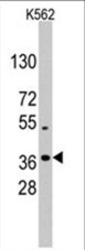
- Western blot analysis of anti-NANOG Antibody (N-term) (AP11497PU-N) in K562 cell line lysates (35 µg/lane). NANOG (arrow) was detected using the purified Pab (1:60 dilution).

- Submitted by
- Acris Antibodies GmbH (provider)
- Main image

- Experimental details
- Western blot analysis of NANOG (arrow) using rabbit polyclonal NANOG Antibody (N-term) (AP11497PU-N). 293 cell lysates (2 µg/lane) either nontransfected (Lane 1) or transiently transfected with the NANOG gene (Lane 2) (Origene Technologies).

- Submitted by
- Acris Antibodies GmbH (provider)
- Main image
- Experimental details
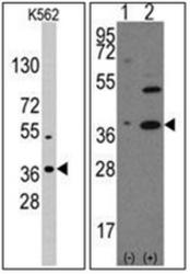
- Left Picture: Western blot analysis of anti-NANOG Antibody (N-term) (Cat.-No AP11497PU-N) in K562 cell line lysates (35 µg/lane). NANOG (arrow) was detected using the purified Pab (1/60 dilution). Right Picture: Western blot analysis of NANOG (arrow) using NANOG Antibody (N-term) (Cat.-No AP11497PU-N). 293 cell lysates (2 ug/lane) either nontransfected (Lane 1) or transiently transfected with the NANOG gene (Lane 2) (Origene Technologies).

Supportive validation
- Submitted by
- Acris Antibodies GmbH (provider)
- Main image

- Experimental details
- Fluorescent confocal image of SY5Y cells stained with NANOG Antibody (N-term) Cat.-No AP11497PU-N. SY5Y cells were fixed with 4% PFA (20 min), permeabilized with Triton X-100 (0.2%, 30 min), then incubated with AP11497PU-N NANOG (N-term) primary antibody (1/500, 2 h at room temperature). For secondary antibody, Alexa Fluor® 488 conjugated donkey anti-rabbit antibody (green) was used (1/1000, 1h). Cytoplasmic actin was counterstained with Alexa Fluor® 555 (red) conjugated Phalloidin (5.25 μM, 25 min). Nuclei were counterstained with Hoechst 33342 (blue) (10 µg/ml, 3 min). Nanog immunoreactivity is localized mainly to the nuclei of the SY5Y cells.

Supportive validation
- Submitted by
- Acris Antibodies GmbH (provider)
- Main image

- Experimental details
- Formalin-fixed and paraffin-embedded human Spleen tissue reacted with NANOG Antibody (N-term) Cat.-No AP11497PU-N, which was peroxidase-conjugated to the secondary antibody, followed by AEC staining. This data demonstrates the use of this antibody for immunohistochemistry; clinical relevance has not been evaluated.

Supportive validation
- Submitted by
- Acris Antibodies GmbH (provider)
- Main image
- Experimental details
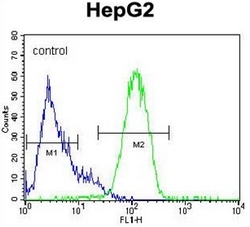
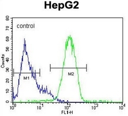
- Flow cytometric analysis of HepG2 cells using NANOG Antibody (N-term) (Cat.-No AP11497PU-N) (right histogram) compared to a negative control cell (left histogram).FITC-conjugated goat-anti-rabbit secondary antibodies were used for the analysis.