 Explore
 Validate
 Learn
Learn Western blot
Antibody data
- Antibody Data
- Antigen structure
- References [2]
- Comments [0]
- Validations
- Western blot [1]
- Immunocytochemistry [1]
- Immunohistochemistry [1]
Submit
Validation data
Reference
Comment
Report error
- Product number
- MA5-14882 - Provider product page

- Provider
- Invitrogen Antibodies
- Product name
- FAS Monoclonal Antibody (H.831.6)
- Antibody type
- Monoclonal
- Antigen
- Synthetic peptide
- Description
- It is not recommended to aliquot this antibody.
- Reactivity
- Human, Rat
- Host
- Rabbit
- Isotype
- IgG
- Antibody clone number
- H.831.6
- Vial size
- 100 µL
- Concentration
- 206 µg/mL
- Storage
- -20°C
Submitted references Mechanical Study of Jian-Gan-Xiao-Zhi Decoction on Nonalcoholic Fatty Liver Disease Based on Integrated Network Pharmacology and Untargeted Metabolomics.
Immunohistological analysis of cell cycle and apoptosis regulators in thymus.
Cao YJ, Li HZ, Zhao J, Sun YM, Jin XW, Lv SQ, Luo JY, Fang XX, Wen WB, Liao JB
Evidence-based complementary and alternative medicine : eCAM 2022;2022:2264394
Evidence-based complementary and alternative medicine : eCAM 2022;2022:2264394
Immunohistological analysis of cell cycle and apoptosis regulators in thymus.
Bai M, Doukas M, Papoudou-Bai A, Barbouti A, Stefanaki K, Galani V, Kanavaros P
Annals of anatomy = Anatomischer Anzeiger : official organ of the Anatomische Gesellschaft 2013 Mar;195(2):159-65
Annals of anatomy = Anatomischer Anzeiger : official organ of the Anatomische Gesellschaft 2013 Mar;195(2):159-65
No comments: Submit comment
Supportive validation
- Submitted by
- Invitrogen Antibodies (provider)
- Main image
- Experimental details
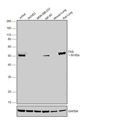
- Western Blot was performed using Anti-FAS Monoclonal Antibody (H.831.6) (Product # MA5-14882) and ~ 50 kDa band corresponding to glycosylated FAS (Tumor necrosis factor receptor superfamily member 6) was observed in Jurkat, NK-92 and rat lung tissue but not in HUVEC or MDA-MB-231 which are reported low expressing for FAS. Whole cell extracts (30 µg lysate) of Jurkat (Lane 1), HUVEC (Lane 2), MDA-MB-231 (Lane 3), NK-92 (Lane 4) and tissue extracts (30 µg lysate) of Mouse Lung (Lane 5) and Rat Lung (Lane 6) were electrophoresed using NuPAGE™ 10% Bis-Tris Protein Gel (Product # NP0301BOX). Resolved proteins were then transferred onto a nitrocellulose membrane (Product # IB23002) by iBlot® 2 Dry Blotting System (Product # IB21001). The blot was probed with the primary antibody (1:1000 dilution) and detected by chemiluminescence with Goat anti-Rabbit IgG (H+L) Superclonal™ Recombinant Secondary Antibody, HRP (Product # A27036, 1:10000 dilution) using the iBright FL 1000 (Product # A32752). Chemiluminescent detection was performed using Novex® ECL Chemiluminescent Substrate Reagent Kit (Product # WP20005).

Supportive validation
- Submitted by
- Invitrogen Antibodies (provider)
- Main image
- Experimental details
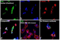
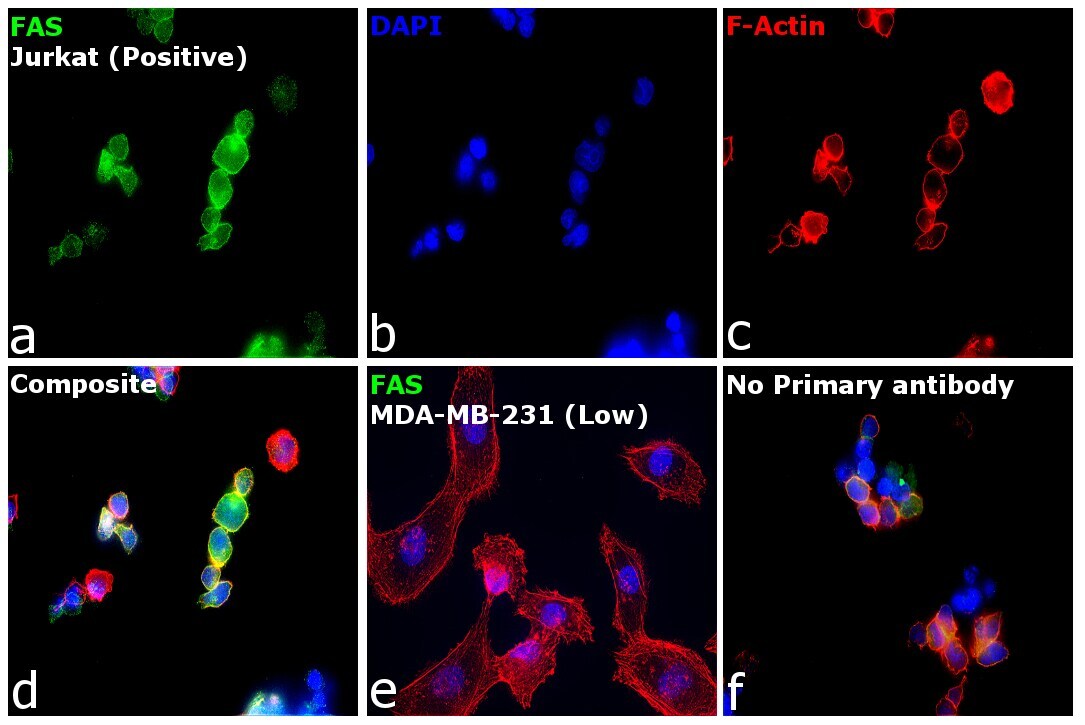
- Immunofluorescence analysis of FAS (Tumor necrosis factor receptor superfamily member 6) was performed using Jurkat and MDA-MB-231 cells. The cells were fixed with 4% paraformaldehyde for 10 minutes, permeabilized with 0.1% Triton™ X-100 for 10 minutes, and blocked with 2% BSA for 1 hour at room temperature. The cells were labeled with FAS Monoclonal Antibody (H.831.6) (Product # MA5-14882) at 1:100 in 0.1% BSA, incubated at 4 degree celsius overnight and then labeled with Donkey anti-Rabbit IgG (H+L) Highly Cross-Adsorbed Secondary Antibody, Alexa Fluor Plus 488 (Product # A32790), (1:2500), for 45 minutes at room temperature (Panel a: Green). Nuclei (Panel b: Blue) were stained with ProLong™ Diamond Antifade Mountant with DAPI (Product # P36962). F-actin (Panel c: Red) was stained with Rhodamine Phalloidin (Product # R415, 1:300). Panel d represents the merged image of Jurkat cells showing membrane localization of FAS while panel e represents MDA-MB-231 cells with no signal for the same. Panel f represents control Jurkat cells with no primary antibody to assess background. The images were captured at 60X magnification with EVOS™ M7000 Imaging System (Product # AMF7000).
Supportive validation
- Submitted by
- Invitrogen Antibodies (provider)
- Main image

- Experimental details
- Immunohistochemical analysis of Fas in paraffin embedded human colon using a Fas monoclonal antibody (Product # MA5-14882) in the presence of control peptide (left) or antigen specific peptide (right).
