 Explore
 Validate
 Learn
Learn Western blot
Antibody data
- Antibody Data
- Antigen structure
- References [0]
- Comments [0]
- Validations
- Western blot [1]
- Flow cytometry [1]
Submit
Validation data
Reference
Comment
Report error
- Product number
- AF6124 - Provider product page

- Provider
- Novus Biologicals
- Product name
- Sheep Polyclonal FOLR4 Antibody
- Antibody type
- Polyclonal
- Description
- Immunogen affinity purified. Detects mouse FOLR4 in direct ELISAs and Western blots. In direct ELISAs, less than 1% cross-reactivity with recombinant human (rh) FOLR1, rhFOLR2, rhFOLR3, and rhFOLR4 is observed.
- Reactivity
- Mouse
- Host
- Sheep
- Isotype
- IgG
- Vial size
- 100 ug
- Concentration
- LYOPH
- Storage
- Use a manual defrost freezer and avoid repeated freeze-thaw cycles. 12 months from date of receipt, -20 to -70 degreesC as supplied. 1 month, 2 to 8 degreesC under sterile conditions after reconstitution. 6 months, -20 to -70 degreesC under sterile conditions after reconstitution.
No comments: Submit comment
Supportive validation
- Submitted by
- Novus Biologicals (provider)
- Main image

- Experimental details
- Detection of Mouse FOLR4 by Western Blot. Western blot shows lysates of mouse CD+CD25+ regulatory T cells. PVDF Membrane was probed with 1 µg/mL of Sheep Anti-Mouse FOLR4 Antigen Affinity-purified Polyclonal Antibody (Catalog # AF6124) followed by HRP-conjugated Anti-Sheep IgG Secondary Antibody (Catalog # HAF016). A specific band was detected for FOLR4 at approximately 37 kDa (as indicated). This experiment was conducted under reducing conditions and using Immunoblot Buffer Group 8.

Supportive validation
- Submitted by
- Novus Biologicals (provider)
- Main image
- Experimental details
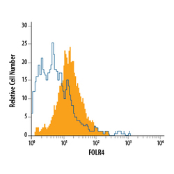
- Detection of FOLR4 in Mouse Splenocytes Gated on FoxP3 Cells by Flow Cytometry. Mouse splenocytes gated on FoxP3 cells treated with 10 μL/mL anti-CD3/anti-CD28, Recombinant Mouse IL-2 (Catalog # 402-ML), and Human TGF-beta 1 (Catalog # 100-B) for 3 days to induce T regulatory cells (Tregs) were stained with Sheep Anti-Mouse FOLR4 Antigen Affinity-purified Polyclonal Antibody (Catalog # AF6124, filled histogram) or isotype control antibody (Catalog # 5-001-A, open histogram), followed by Allophycocyanin-conjugated Anti-Sheep IgG Secondary Antibody (Catalog # F0127). To facilitate intracellular staining, cells were fixed and permeabilized with Flow Cytometry FoxP3 Staining Buffer (Catalog # FC011).
