 Explore
 Validate
 Learn
Learn Western blot
Antibody data
- Antibody Data
- Antigen structure
- References [1]
- Comments [0]
- Validations
- Western blot [1]
- Flow cytometry [1]
- Chromatin Immunoprecipitation [1]
Submit
Validation data
Reference
Comment
Report error
- Product number
- MAB33341 - Provider product page

- Provider
- R&D Systems
- Product name
- Human BMI-1 Antibody
- Antibody type
- Monoclonal
- Description
- Protein A or G purified from hybridoma culture supernatant. Detects human BMI-1 in direct ELISAs and Western blots.
- Reactivity
- Human
- Host
- Mouse
- Conjugate
- Unconjugated
- Antigen sequence
P35226- Isotype
- IgG
- Antibody clone number
- 384515
- Vial size
- 50 ug
- Concentration
- LYOPH
- Storage
- Use a manual defrost freezer and avoid repeated freeze-thaw cycles. 12 months from date of receipt, -20 to -70 °C as supplied. 1 month, 2 to 8 °C under sterile conditions after reconstitution. 6 months, -20 to -70 °C under sterile conditions after reconstitution.
Submitted references BMI1 reprogrammes histone acetylation and enhances c-fos pathway via directly binding to Zmym3 in malignant myeloid progression.
Shen H, Chen Z, Ding X, Qi X, Cen J, Wang Y, Yao L, Chen Y
Journal of cellular and molecular medicine 2014 Jun;18(6):1004-17
Journal of cellular and molecular medicine 2014 Jun;18(6):1004-17
No comments: Submit comment
Supportive validation
- Submitted by
- R&D Systems (provider)
- Main image

- Experimental details
- Detection of Human BMI-1 by Western Blot. Western blot shows lysates of HeLa human cervical epithelial carcinoma cell line. Gels were loaded with 30 μg of whole cell lysate (WCL), 20 μg of cytoplasmic (Cyto), and 10 μg of nuclear extracts (Nuc). PVDF membrane was probed with 0.1 µg/mL Mouse Anti-Human BMI-1 Monoclonal Antibody (Catalog # MAB33341) followed by HRP-conjugated Anti-Mouse IgG Secondary Antibody (Catalog # HAF007). A specific band for BMI-1 was detected at approximately 45 kDa (as indicated). This experiment was conducted under reducing conditions and using Immunoblot Buffer Group 4.

Supportive validation
- Submitted by
- R&D Systems (provider)
- Main image

- Experimental details
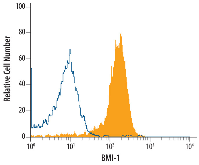
- Detection of BMI-1 in HeLa Human Cell Line by Flow Cytometry. HeLa human cervical epithelial carcinoma cell line was stained with Mouse Anti-Human BMI-1 Monoclonal Antibody (Catalog # MAB33341, filled histogram) or isotype control antibody (Catalog # MAB003, open histogram), followed by Phycoerythrin-conjugated Anti-Mouse IgG F(ab')2 Secondary Antibody (Catalog # F0102B). To facilitate intracellular staining, cells were fixed with paraformaldehyde and permeabilized with saponin.
Supportive validation
- Submitted by
- R&D Systems (provider)
- Main image

- Experimental details
- Detection of BMI-1-regulated Genes by Chromatin Immunoprecipitation. HeLa human cervical epithelial carcinoma cell line was fixed using formaldehyde, resuspended in lysis buffer, and sonicated to shear chromatin. BMI-1/DNA complexes were immunoprecipitated using 5 μg Mouse Anti-Human BMI-1 Monoclonal Antibody (Catalog # MAB33341) or control antibody (Catalog # MAB003) for 15 minutes in an ultrasonic bath, followed by Biotinylated Anti-Mouse IgG Secondary Antibody (Catalog # BAF007). Immunocomplexes were captured using 50 μL of MagCellect Streptavidin Ferrofluid (Catalog # MAG999) and DNA was purified using chelating resin solution. The hoxc13 promoter was detected by standard PCR.
