Antibody data
- Antibody Data
- Antigen structure
- References [6]
- Comments [0]
- Validations
- Western blot [3]
- Immunocytochemistry [1]
- Flow cytometry [1]
- Other assay [2]
Submit
Validation data
Reference
Comment
Report error
- Product number
- MA5-11291 - Provider product page

- Provider
- Invitrogen Antibodies
- Product name
- CDK5 Antibody Cocktail
- Antibody type
- Monoclonal
- Antigen
- Recombinant full-length protein
- Description
- MA5-11291 targets Cdk5 in IF, IP, and WB applications and shows reactivity with Human, mouse, and Rat samples.
- Antibody clone number
- DC17, DC34
- Concentration
- 0.2 mg/mL
Submitted references Cyclin-dependent kinase-5 and p35/p25 activators in schizophrenia and major depression prefrontal cortex: basal contents and effects of psychotropic medications.
The protective effects of tanshinone IIA on neurotoxicity induced by β-amyloid protein through calpain and the p35/Cdk5 pathway in primary cortical neurons.
Crosstalk between cdk5 and MEK-ERK signalling upon opioid receptor stimulation leads to upregulation of activator p25 and MEK1 inhibition in rat brain.
Implication of cyclin-dependent kinase 5 in the neuroprotective properties of lithium.
(+/-)-huprine Y, (-)-huperzine A and tacrine do not show neuroprotective properties in an apoptotic model of neuronal cytoskeletal alteration.
(+/-)-huprine Y, (-)-huperzine A and tacrine do not show neuroprotective properties in an apoptotic model of neuronal cytoskeletal alteration.
Ramos-Miguel A, Meana JJ, García-Sevilla JA
The international journal of neuropsychopharmacology 2013 Apr;16(3):683-9
The international journal of neuropsychopharmacology 2013 Apr;16(3):683-9
The protective effects of tanshinone IIA on neurotoxicity induced by β-amyloid protein through calpain and the p35/Cdk5 pathway in primary cortical neurons.
Shi LL, Yang WN, Chen XL, Zhang JS, Yang PB, Hu XD, Han H, Qian YH, Liu Y
Neurochemistry international 2012 Jul;61(2):227-35
Neurochemistry international 2012 Jul;61(2):227-35
Crosstalk between cdk5 and MEK-ERK signalling upon opioid receptor stimulation leads to upregulation of activator p25 and MEK1 inhibition in rat brain.
Ramos-Miguel A, García-Sevilla JA
Neuroscience 2012 Jul 26;215:17-30
Neuroscience 2012 Jul 26;215:17-30
Implication of cyclin-dependent kinase 5 in the neuroprotective properties of lithium.
Jordà EG, Verdaguer E, Canudas AM, Jiménez A, Garcia de Arriba S, Allgaier C, Pallàs M, Camins A
Neuroscience 2005;134(3):1001-11
Neuroscience 2005;134(3):1001-11
(+/-)-huprine Y, (-)-huperzine A and tacrine do not show neuroprotective properties in an apoptotic model of neuronal cytoskeletal alteration.
Jordá EG, Verdaguer E, Jiménez A, Canudas AM, Rimbau V, Camps P, Muñoz-Torrero D, Camins A, Pallàs M
Journal of Alzheimer's disease : JAD 2004 Dec;6(6):577-83; discussion 673-81
Journal of Alzheimer's disease : JAD 2004 Dec;6(6):577-83; discussion 673-81
No comments: Submit comment
Supportive validation
- Submitted by
- Invitrogen Antibodies (provider)
- Main image

- Experimental details
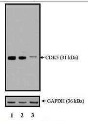
- Western blot analysis of CDK5 was performed with 10 µg of HeLa cells transfected with Transfection Reagent alone (Lane 1), 100nM Non-Targeting control siRNA (Lane 2), or 100nM siRNA against CDK5 (Lane 3). Proteins were resolved using a NuPAGE® Novex 4-12% Bis-Tris Gel (Product # NP0322BOX), XCell SureLock™ Electrophoresis System (Product # EI0002), and a protein size ladder. Proteins were wet transferred to a Pierce Nitrocellulose Membrane (Product # 88025) OR Pierce PVDF Membrane (Product # 88518) and blocked with Pierce Starting Block T20 (PBS) Blocking Buffer (Product # 37539) for 1 hour at room temperature. CDK5 was detected at ~ 31 kDa using CDK5 Mouse monoclonal antibody (Product # MA5-11291) diluted in Pierce Starting Block T20 (PBS) Blocking Buffer 4°C overnight on a rocking platform. Pierce Goat Anti-Mouse (Product # 31437) HRP-Conjugated Antibodies at a 1:2500 dilution were used and chemiluminescent detection was performed using Pierce Supersignal West Dura Maximum Sensitivity Substrate (Product # 37071). Relative density of the bands normalized to GAPDH (36 kDa). CDK5 Antibody (Product # MA5-11291) confirms silencing of CDK5 expression.
- Submitted by
- Invitrogen Antibodies (provider)
- Main image

- Experimental details
- Western blot of Cdk5 using Cdk5 Monoclonal Antibody (Product # MA5-11291) on IMR-5 Cells.
- Submitted by
- Invitrogen Antibodies (provider)
- Main image

- Experimental details
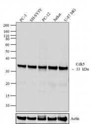
- Western blot analysis was performed on whole cell extracts (30 µg lysate) of PC-3 (lane 1), SH-SY5Y (lane 2), PC-12 (lane 3), Jurkat (lane 4) and U-87 MG (lane 5). The blots were probed with Anti-Cdk5 Mouse Monoclonal Antibody (Product # MA5-11291, 1-2 µg/mL) and detected by chemiluminescence oat anti-Mouse IgG (H+L) Secondary Antibody, HRP conjugate (Product # 62-6520, 1:4000 dilution). A 33 kDa band corresponding to Cdk5 was observed across cell lines tested. Known quantity of protein samples were electrophoresed using Novex® NuPAGE® 4-12 % Bis-Tris gel (Product # NP0321BOX), XCell SureLock™ Electrophoresis System (Product # EI0002) and Novex® Sharp Pre-Stained Protein Standard (Product # LC5800). Resolved proteins were then transferred onto a nitrocellulose membrane with iBlot® 2 Dry Blotting System (Product # IB21001). The membrane was probed with the relevant primary and secondary Antibody using iBind™ Flex Western Starter Kit (Product # SLF2000S). Chemiluminescent detection was performed using Pierce™ ECL Western Blotting Substrate (Product # 32106).
Supportive validation
- Submitted by
- Invitrogen Antibodies (provider)
- Main image

- Experimental details
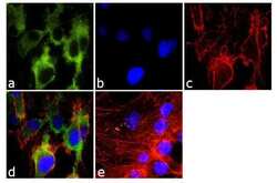
- Immunofluorescence analysis of Cdk5 was done on 70% confluent log phase MCF-7 cells. The cells were fixed with 4% paraformaldehyde for 10 minutes, permeabilized with 0.1% Triton™ X-100 for 10 minutes, and blocked with 1% BSA for 1 hour at room temperature. The cells were labeled with Cdk5 (DC17 + DC34) Mouse Monoclonal Antibody (Product # MA5-11291) at 2 µg/mL in 0.1% BSA and incubated for 3 hours at room temperature and then labeled with Goat anti-Mouse IgG (H+L) Superclonal™ Secondary Antibody, Alexa Fluor® 488 conjugate (Product # A28175) at a dilution of 1:2000 for 45 minutes at room temperature (Panel a: green). Nuclei (Panel b: blue) were stained with SlowFade® Gold Antifade Mountant with DAPI (Product # S36938). F-actin (Panel c: red) was stained with Alexa Fluor® 555 Rhodamine Phalloidin (Product # R415, 1:300). Panel d is a merged image showing cytoplasmic localization. Panel e is a no primary antibody control. The images were captured at 60X magnification.
Supportive validation
- Submitted by
- Invitrogen Antibodies (provider)
- Main image

- Experimental details
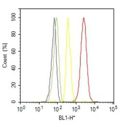
- Flow cytometry analysis of Cdk5 was done on PC-3 cells. Cells were fixed with 70% ethanol for 10 minutes, permeabilized with 0.25% Triton™ X-100 for 20 minutes, and blocked with 5% BSA for 30 minutes at room temperature. Cells were labeled with Cdk5 Mouse Monoclonal Antibody (MA511291, red histogram) or with mouse isotype control (yellow histogram) at 3-5 ug/million cells in 2.5% BSA. After incubation at room temperature for 2 hours, the cells were labeled with Alexa Fluor® 488 Rabbit Anti-Mouse Secondary Antibody (A11059) at a dilution of 1:400 for 30 minutes at room temperature. The representative 10,000 cells were acquired and analyzed for each sample using an Attune® Acoustic Focusing Cytometer. The purple histogram represents unstained control cells and the green histogram represents no-primary-antibody control.
Supportive validation
- Submitted by
- Invitrogen Antibodies (provider)
- Main image

- Experimental details
- Immunoprecipitation of Cdk5 using Cdk5 Monoclonal Antibody (Product # MA5-11291) on Native Human LS174T Cells.
- Submitted by
- Invitrogen Antibodies (provider)
- Main image

- Experimental details
- Immunoprecipitation and Western blot of Cdk5 was performed using Jurkat cell lysates. Cells were lysed using ice cold IP lysis/wash buffer and pre-cleared using 50 mL of bead slurry per mL of cell lysate. Antigen-antibody complexes were formed by incubating 0.5 mL pre-cleared cell lysate on ice for 3hrs with 8-15 µg of Cdk5 monoclonal antibody (Product # MA5-11291) crosslinked to Protein A/G plus agarose. The immune complexes were eluted using 60 µL sample buffer boiled at 95§C for 5 min and loaded onto an SDS-PAGE gel; Input (lane 1), Jurkat IP (Lane 2) and Jurkat supernatant (lane 3). The membrane was probed with a Cdk5 monoclonal antibody (Product # MA5-11291) at a dilution of 6 µg/mg of lysate followed by detection using an HRP-conjugated goat anti-mouse IgG + IgM (H+L) cross-adsorbed secondary antibody. Chemiluminescent detection was performed using an exposure time of 20s, resulting in a ~30 kDa band on input and IP lysates.
 Explore
 Validate
 Learn
Learn Western blot