 Explore
 Validate
 Learn
Learn Western blot
 ELISA
ELISAAntibody data
- Antibody Data
- Antigen structure
- References [1]
- Comments [0]
- Validations
- Western blot [2]
- Immunocytochemistry [1]
- Immunohistochemistry [3]
- Flow cytometry [3]
- Chromatin Immunoprecipitation [1]
- Other assay [2]
Submit
Validation data
Reference
Comment
Report error
- Product number
- 700031 - Provider product page

- Provider
- Invitrogen Antibodies
- Product name
- Phospho-JNK1/JNK2 (Thr183, Tyr185) Recombinant Rabbit Monoclonal Antibody (D12H7L17)
- Antibody type
- Monoclonal
- Antigen
- Synthetic peptide
- Reactivity
- Human, Mouse
- Host
- Rabbit
- Isotype
- IgG
- Antibody clone number
- D12H7L17
- Vial size
- 100 µg
- Concentration
- 0.5 mg/mL
- Storage
- Store at 4°C short term. For long term storage, store at -20°C, avoiding freeze/thaw cycles.
Submitted references Tumor necrosis factor-alpha mediates activation of NF-κB and JNK signaling cascades in retinal ganglion cells and astrocytes in opposite ways.
Dvoriantchikova G, Ivanov D
The European journal of neuroscience 2014 Oct;40(8):3171-8
The European journal of neuroscience 2014 Oct;40(8):3171-8
No comments: Submit comment
Supportive validation
- Submitted by
- Invitrogen Antibodies (provider)
- Main image

- Experimental details
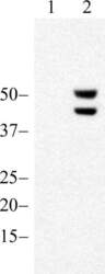
- Western blot analysis of JNK1/2 (pT183/pY185) was performed by loading 20 µg of MCF7 (lane1) and MCF7 treated for 30 minutes with 25 µg/mL of Anisomycin cell lysate using NuPAGE® Novex® 10% Bis-Tris gel (Product # NP0301BOX), XCell SureLock Electrophoresis System (Product # EI0002), Novex® Sharp Pre-Stained Protein Standard (Product # LC5800), and iBlot® Dry Blotting System (Product # IB21001). Proteins were transferred to a nitrocellulose membrane and blocked with 5% skim milk for 1 hour at room temperature. JNK1/2 (pT183/pY185) was detected at ~46 and 54 kDa using JNK1/2 (pT183/pY185) Recombinant Rabbit Monoclonal Antibody (Product # 700031) at 0.5-1 µg/mL in 2.5% skim milk at 4°C overnight on a rocking platform. Goat anti-Rabbit IgG - HRP Secondary Antibody (Product # G-21234) at 1:5000 dilution was used and chemiluminescent detection was performed using Pierce™ ECL Western blotting Substrate (Product # 32106).

- Submitted by
- Invitrogen Antibodies (provider)
- Main image
- Experimental details
- Western blot analysis of Phospho-JNK1/JNK2 pThr183+pTyr185 in untreated HEK293 lysates (lane 1) or Anisomycin-treated HEK293 lysates (lane 2) using a Phospho-JNK1/JNK2 pThr183+pTyr185 recombinant rabbit monoclonal antibody (Product # 700031) at a dilution of 0.1 µg/mL.

Supportive validation
- Submitted by
- Invitrogen Antibodies (provider)
- Main image

- Experimental details
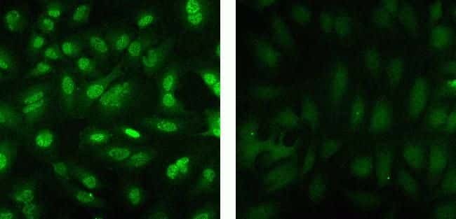
- Immunofluorescent analysis of Phospho-JNK1/JNK2 pThr183+pTyr185 in HeLa cells using a Phospho-JNK1/JNK2 pThr183+pTyr185 recombinant rabbit monoclonal antibody (Product # 700031) at a dilution of 5 µg/mL in the absence of peptide (left) or in the presence of the immunogenic peptide (right), followed by detection using an Alexa Fluor 488-conjugated goat anti-rabbit secondary antibody at a dilution of 1:1000.
Supportive validation
- Submitted by
- Invitrogen Antibodies (provider)
- Main image
- Experimental details
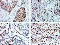
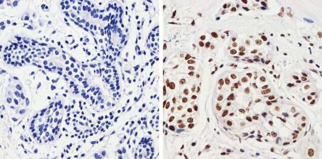
- Immunohistochemistry analysis of Phospho-JNK1/JNK2 pThr183/pTyr185 in formalin-fixed, paraffin-embedded human squamous lung (top left), breast (top right), gastric carninoma (bottom left) and mesothelioma tissue (bottom right) using a Phospho-JNK1/JNK2 pThr183/pTyr185 monoclonal antibody (Product # 700031) at a dilution of 2 µg/mL. Tissues were pretreated with EDTA and staining was visualized using DAB. Images were taken at a magnification of 40x. Results show nuclear staining in tumor cells.

- Submitted by
- Invitrogen Antibodies (provider)
- Main image
- Experimental details
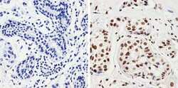
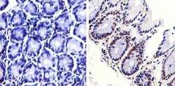
- Immunohistochemistry analysis of JNK1/2 (pT183/pY185) showing staining in the nucleus of paraffin-embedded human breast carcinoma (right) compared to a negative control without primary antibody (left). To expose target proteins, antigen retrieval was performed using 10 mM sodium citrate (pH 6.0), microwaved for 8-15 min. Following antigen retrieval, tissues were blocked in 3% H2O2-methanol for 15 min at room temperature, washed with ddH2O and PBS, and then probed with JNK1/2 (pT183/pY185) Monoclonal antibody (Product # 700031) diluted in 3% BSA-PBS at a dilution of 1:50 overnight at 4°C in a humidified chamber. Tissues were washed extensively in PBST and detection was performed using a HRP-conjugated secondary antibody followed by colorimetric detection using a DAB kit. Tissues were counterstained with hematoxylin and dehydrated with ethanol and xylene to prep for mounting.
- Submitted by
- Invitrogen Antibodies (provider)
- Main image
- Experimental details
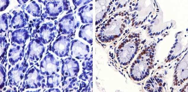
- Immunohistochemistry analysis of JNK1/2 (pT183/pY185) showing staining in the nucleus of paraffin-embedded mouse colon tissue (right) compared to a negative control without primary antibody (left). To expose target proteins, antigen retrieval was performed using 10 mM sodium citrate (pH 6.0), microwaved for 8-15 min. Following antigen retrieval, tissues were blocked in 3% H2O2-methanol for 15 min at room temperature, washed with ddH2O and PBS, and then probed with JNK1/2 (pT183/pY185) Monoclonal antibody (Product # 700031) diluted in 3% BSA-PBS at a dilution of 1:20 overnight at 4°C in a humidified chamber. Tissues were washed extensively in PBST and detection was performed using a HRP-conjugated secondary antibody followed by colorimetric detection using a DAB kit. Tissues were counterstained with hematoxylin and dehydrated with ethanol and xylene to prep for mounting.
Supportive validation
- Submitted by
- Invitrogen Antibodies (provider)
- Main image
- Experimental details
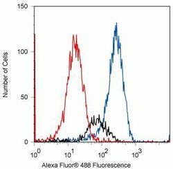
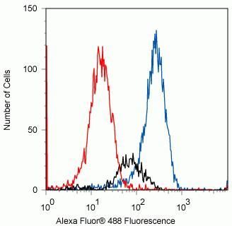
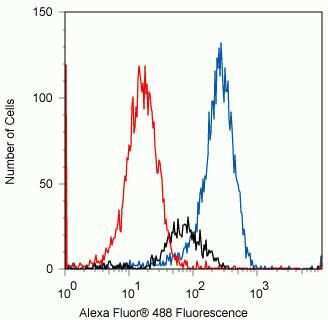
- Flow cytometry analysis of Phospho-JNK1 pThr183/pTyr185 in Jurkat cells stimulated with 25 µg/mL anisomycin for 45 min using a Phospho-JNK1 pThr183/pTyr185 recombinant rabbit monoclonal antibody (Product # 700031) at a dilution of 0.5 µg. Cells were fixed and permeabilized using FIX & PERM (Product # GAS-004) reagent, and detection was performed using an Alexa Fluor 488 goat anti-rabbit IgG (red) compared to a control without primary antibody (blue) and unstimulated cells (black).
- Submitted by
- Invitrogen Antibodies (provider)
- Main image
- Experimental details
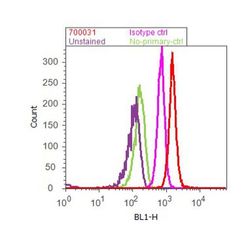
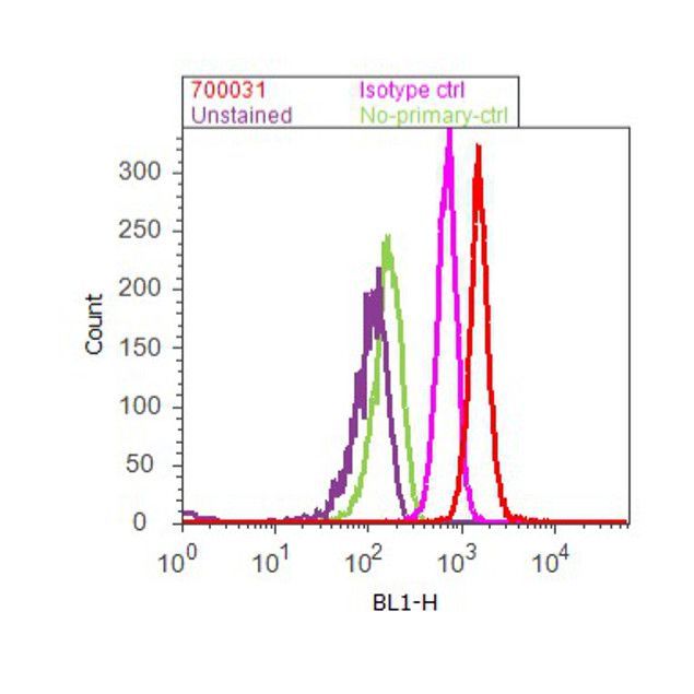
- Flow cytometry analysis of JNK1/2 [pT183/pY185] was done on serum starved MCF7 cells. Cells were fixed with 70% ethanol for 10 minutes, permeabilized with 0.25% Triton™ X-100 for 20 minutes, and blocked with 5% BSA for 30 minutes at room temperature. Cells were labeled with ABfinity™ JNK1/2 [pT183/pY185] Recombinant Rabbit Monoclonal Antibody (700031, red histogram) or with rabbit isotype control (pink histogram) at 3-5 µg/million cells in 2.5% BSA. After incubation at room temperature for 2 hours, the cells were labeled with Alexa Fluor® 488 Goat Anti-Rabbit Secondary Antibody (A11008) at a dilution of 1:400 for 30 minutes at room temperature. The representative 10,000 cells were acquired and analyzed for each sample using an Attune® Acoustic Focusing Cytometer. The purple histogram represents unstained control cells and the green histogram represents no-primary-antibody control.
- Submitted by
- Invitrogen Antibodies (provider)
- Main image
- Experimental details
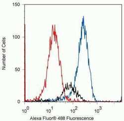
- Flow cytometry analysis of Phospho-JNK1 pThr183/pTyr185 in Jurkat cells stimulated with 25 µg/mL anisomycin for 45 min using a Phospho-JNK1 pThr183/pTyr185 recombinant rabbit monoclonal antibody (Product # 700031) at a dilution of 0.5 µg. Cells were fixed and permeabilized using FIX & PERM (Product # GAS-004) reagent, and detection was performed using an Alexa Fluor 488 goat anti-rabbit IgG (red) compared to a control without primary antibody (blue) and unstimulated cells (black).
Supportive validation
- Submitted by
- Invitrogen Antibodies (provider)
- Main image

- Experimental details
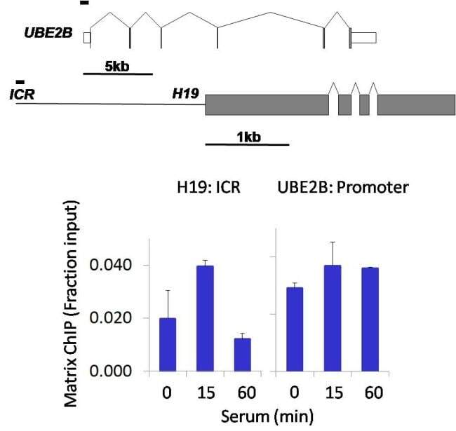
- Chromatin immunoprecipitation analysis of Phospho-JNK1+JNK2 (pThr183+pTyr185) was performed using cross-linked chromatin from 1 x 10^6 HCT116 human colon carcinoma cells treated with serum for 0, 15, and 60 minutes. Immunoprecipitation was performed using a multiplex microplate Matrix ChIP assay (see reference for Matrix ChIP protocol: http://www.ncbi.nlm.nih.gov/pubmed/22098709) with 1.0 µL/100 µL well volume of a Phospho-JNK1+JNK2 (pThr183+pTyr185) rabbit monoclonal antibody (Product # 700031). Chromatin aliquots from ~1 x 10^5 cells were used per ChIP pull-down. Quantitative PCR data were done in quadruplicate using 1 µL of eluted DNA in 2 µL SYBR real-time PCR reactions containing primers to amplify the promoter region of human UBE2B, or the imprinting control region (ICR) of the human H19 locus. PCR calibration curves were generated for each primer pair from a dilution series of sheared total genomic DNA. Quantitation of immunoprecipitated chromatin is presented as signal relative to the total amount of input chromatin. Results represent the mean +/- SEM for three experiments. A schematic representation of the human UBE2B and H19 loci are shown above the data where boxes represent exons (grey boxes = translated regions, white boxes = untranslated regions), the zigzag lines represent introns, and the straight line represents upstream sequence. Regions amplified by UBE2B and H19 primers are represented by black bars. Data courtesy of the Innovators Program.
Supportive validation
- Submitted by
- Invitrogen Antibodies (provider)
- Main image
- Experimental details
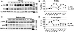
- Figure 4 Representative (A and C) Western blots and (B and D) densitometric analysis of JNK (total) and phosphorylated JNK (p-JNK) protein levels in the TNF-treated primary RGCs and astrocytes revealed sustained JNK activation in TNF-treated RGCs, while primary astrocytes demonstrated transient JNK activation. Whole-cell extracts (25 mug) were collected from zero to 90 min as well as 2, 3 and 24 h after TNF treatment, and the JNK state was tested by Western blot analysis using an antibody against JNK and p-JNK. The JNK and p-JNK intensities were normalised to GAPDH and the results expressed as percentages of the corresponding values in the cells treated for 0 min (0 m). Values are means +- SEM; n = 3.

- Submitted by
- Invitrogen Antibodies (provider)
- Main image
- Experimental details
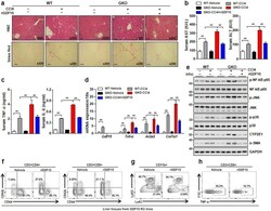
- Figure 6 Recombinant GDF15 ameliorates CCl 4 -induced liver inflammation and fibrosis. WT and GDF15 KO (GKO) mice (n = 5/group) were treated with CCl 4 (2 mL/kg in olive oil, 20% v/v) and recombinant GDF15 (rGDF15, 0.5 mg/kg). ( a ) H&E and Sirius Red staining of liver sections (original magnification, x 200; bars, 100 mum). ( b ) Serum levels of AST and ALT. ( c ) Serum levels of TNF-alpha and IL-6. ( d ) Real-time PCR analysis of hepatic Gdf15 , Tnf-alpha , Acta2 , and Col1a1 . ( e ) Western blots of liver tissue. ( f ) Percentages of hepatic CD4 + CD44 + and CD8 + CD44 + T cells in GDF15 KO mice treated with vehicle or rGDF15. ( g ) Percentages of hepatic monocytes (CD11b + Ly6C high ) and neutrophils (CD11b + Ly6G + ) in GDF15 KO mice treated with vehicle or rGDF15. ( h ) Intracellular staining for TNF-alpha in CD8 + T cells in the livers of GDF15 KO mice treated with vehicle or rGDF15. All data are expressed as the mean +- SEM. **P < 0.01, versus the corresponding controls.
