Explore
 Validate
 Learn
Learn Western blot
Antibody data
- Antibody Data
- Antigen structure
- References [5]
- Comments [0]
- Validations
- Western blot [1]
- Immunocytochemistry [1]
- Immunohistochemistry [1]
Submit
Validation data
Reference
Comment
Report error
- Product number
- AF2957 - Provider product page

- Provider
- R&D Systems
- Product name
- Human S100P Antibody
- Antibody type
- Polyclonal
- Description
- Antigen Affinity-purified. Detects human S100P in direct ELISAs and Western blots. In Western blots, less than 5% cross-reactivity with recombinant human (rh) S100B and rhS100A10 is observed.
- Reactivity
- Human
- Host
- Goat
- Conjugate
- Unconjugated
- Antigen sequence
P25815- Isotype
- IgG
- Vial size
- 100 ug
- Concentration
- LYOPH
- Storage
- Use a manual defrost freezer and avoid repeated freeze-thaw cycles. 12 months from date of receipt, -20 to -70 °C as supplied. 1 month, 2 to 8 °C under sterile conditions after reconstitution. 6 months, -20 to -70 °C under sterile conditions after reconstitution.
Submitted references Morphological subclassification of intrahepatic cholangiocarcinoma: etiological, clinicopathological, and molecular features.
Ca2+/S100 proteins act as upstream regulators of the chaperone-associated ubiquitin ligase CHIP (C terminus of Hsc70-interacting protein).
Porcupine expression is associated with the expression of S100P and other cancer-related molecules in non-small cell lung carcinoma.
Porcupine expression is associated with the expression of S100P and other cancer-related molecules in non-small cell lung carcinoma.
Identification and characterization of novel ERC-55 interacting proteins: evidence for the existence of several ERC-55 splicing variants; including the cytosolic ERC-55-C.
Liau JY, Tsai JH, Yuan RH, Chang CN, Lee HJ, Jeng YM
Modern pathology : an official journal of the United States and Canadian Academy of Pathology, Inc 2014 Aug;27(8):1163-73
Modern pathology : an official journal of the United States and Canadian Academy of Pathology, Inc 2014 Aug;27(8):1163-73
Ca2+/S100 proteins act as upstream regulators of the chaperone-associated ubiquitin ligase CHIP (C terminus of Hsc70-interacting protein).
Shimamoto S, Kubota Y, Yamaguchi F, Tokumitsu H, Kobayashi R
The Journal of biological chemistry 2013 Mar 8;288(10):7158-68
The Journal of biological chemistry 2013 Mar 8;288(10):7158-68
Porcupine expression is associated with the expression of S100P and other cancer-related molecules in non-small cell lung carcinoma.
Bartling B, Rehbein G, Simm A, Silber RE, Hofmann HS
International journal of oncology 2010 Apr;36(4):1015-21
International journal of oncology 2010 Apr;36(4):1015-21
Porcupine expression is associated with the expression of S100P and other cancer-related molecules in non-small cell lung carcinoma.
Bartling B, Rehbein G, Simm A, Silber RE, Hofmann HS
International journal of oncology 2010 Apr;36(4):1015-21
International journal of oncology 2010 Apr;36(4):1015-21
Identification and characterization of novel ERC-55 interacting proteins: evidence for the existence of several ERC-55 splicing variants; including the cytosolic ERC-55-C.
Ludvigsen M, Jacobsen C, Maunsbach AB, Honoré B
Proteomics 2009 Dec;9(23):5267-87
Proteomics 2009 Dec;9(23):5267-87
No comments: Submit comment
Supportive validation
- Submitted by
- R&D Systems (provider)
- Main image

- Experimental details
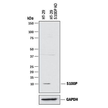
- Detection of Human S100P by Western Blot. Western blot shows lysates of HT-29 human colon adenocarcinoma parental cell line and S100P knockout HT-29 cell line (KO). PVDF membrane was probed with 0.5 µg/mL of Goat Anti-Human S100P Antigen Affinity-purified Polyclonal Antibody (Catalog # AF2957) followed by HRP-conjugated Anti-Goat IgG Secondary Antibody (Catalog # HAF017). A specific band was detected for S100P at approximately 10 kDa (as indicated) in the parental HT-29 cell line, but is not detectable in knockout HT-29 cell line. GAPDH (Catalog # AF5718) is shown as a loading control. This experiment was conducted under reducing conditions and using Immunoblot Buffer Group 1.
Supportive validation
- Submitted by
- R&D Systems (provider)
- Main image

- Experimental details
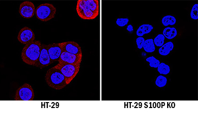
- S100P Specificity is Shown by Immunocytochemistry in Knockout Cell Line. S100P was detected in immersion fixed HT-29 human colon adenocarcinoma cell line but is not detected in S100P knockout (KO) HT-29 Human Cell Line cell line using Goat Anti-Human S100P Antigen Affinity-purified Polyclonal Antibody (Catalog # AF2957) at 0.3 µg/mL for 3 hours at room temperature. Cells were stained using the NorthernLights 557-conjugated Anti-Goat IgG Secondary Antibody (red; Catalog # NL001) and counterstained with DAPI (blue). Specific staining was localized to cytoplasm. View our protocol for Fluorescent ICC Staining of Cells on Coverslips.
Supportive validation
- Submitted by
- R&D Systems (provider)
- Main image

- Experimental details
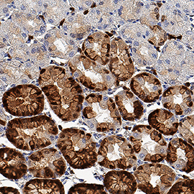
- S100P in Human Stomach. S100P was detected in immersion fixed paraffin-embedded sections of human stomach using Goat Anti-Human S100P Antigen Affinity-purified Polyclonal Antibody (Catalog # AF2957) at 0.3 µg/mL for 1 hour at room temperature followed by incubation with the Anti-Goat IgG VisUCyte™ HRP Polymer Antibody (Catalog # VC004). Tissue was stained using DAB (brown) and counterstained with hematoxylin (blue). Specific staining was localized to nuclei in gastric glands. View our protocol for IHC Staining with VisUCyte HRP Polymer Detection Reagents.