 Explore
 Validate
 Learn
Learn Western blot
Antibody data
- Antibody Data
- Antigen structure
- References [1]
- Comments [0]
- Validations
- Western blot [2]
- Immunocytochemistry [1]
- Immunohistochemistry [3]
- Other assay [1]
Submit
Validation data
Reference
Comment
Report error
- Product number
- PA5-95195 - Provider product page

- Provider
- Invitrogen Antibodies
- Product name
- Ubiquitin B Polyclonal Antibody
- Antibody type
- Polyclonal
- Antigen
- Recombinant full-length protein
- Reactivity
- Human, Mouse, Rat
- Host
- Rabbit
- Isotype
- IgG
- Vial size
- 100 µg
- Concentration
- 500 µg/mL
- Storage
- Store at 4°C short term. For long term storage, store at -20°C, avoiding freeze/thaw cycles.
Submitted references Endogenous Fluorescent Proteins in the Mucus of an Intertidal Polychaeta: Clues for Biotechnology.
Rodrigo AP, Lopes A, Pereira R, Anjo SI, Manadas B, Grosso AR, Baptista PV, Fernandes AR, Costa PM
Marine drugs 2022 Mar 25;20(4)
Marine drugs 2022 Mar 25;20(4)
No comments: Submit comment
Supportive validation
- Submitted by
- Invitrogen Antibodies (provider)
- Main image

- Experimental details
- Western blot analysis of Ubiquitin B in Lane 1: human Caco-2 whole cell lysates, Lane 2: human K562 whole cell lysates, Lane 3: human THP-1 whole cell lysates, Lane 4: rat PC-12 whole cell lysates, Lane 5: rat brain tissue lysates, Lane 6: mouse brain tissue lysates, After Electrophoresis, proteins were transferred to a Nitrocellulose membrane at 150mA for 50-90 minutes. Electrophoresis was performed with 5-20% SDS-PAGE gel (70V, Stacking gel; 90V Resolving gel, Time: 2-3 hours), transferred to a nitrocellulose membrane and blocked using 5% Non-fat Milk/TBS (1.5 hrs at room temperature). Samples were incubated with Ubiquitin B polyclonal antibody (Product # PA5-95195) using a 0.5 µg/mL dilution, followed by a goat anti-rabbit IgG-HRP at a dilution of 1:10,000, and developed with enhanced chemiluminescence (ECL).

- Submitted by
- Invitrogen Antibodies (provider)
- Main image

- Experimental details
- Western blot analysis of Ubiquitin in, Lane 1: human Caco-2 whole cell lysates, Lane 2: human K562 whole cell lysates, Lane 3: human THP-1 whole cell lysates, Lane 4: rat PC-12 whole cell lysates, Lane 5: rat brain tissue lysates, Lane 6: mouse brain tissue lysates, . Electrophoresis was performed on a 5-20% SDS-PAGE gel at 70V (Stacking gel) / 90V (Resolving gel) for 2-3 hours. The sample well of each lane was loaded with 50 µg of sample under reducing conditions. After Electrophoresis, proteins were transferred to a Nitrocellulose membrane at 150mA for 50-90 minutes. The membrane was blocked with 5% Non-fat Milk/ TBS for 1. 5 hour at RT. The membrane was incubated with Ubiquitin B Polyclonal Antibody (Product # PA5-95195) at 0.5 μg/mL overnight at 4°C, then washed with TBS-0. 1% Tween 3 times with 5 minutes each and probed with a goat anti-rabbit IgG-HRP secondary antibody at a dilution of 1:10000 for 1. 5 hour at RT. The signal is developed using an Enhanced Chemiluminescent detection (ECL) kit. A specific band was detected for Ubiquitin at approximately 9KD. The expected band size for Ubiquitin is at 9KD.

Supportive validation
- Submitted by
- Invitrogen Antibodies (provider)
- Main image

- Experimental details
- Immunocytochemistry analysis of Ubiquitin using anti-Ubiquitin antibody (Product # PA5-95195) . Ubiquitin was detected in a section of A431 cells. Enzyme antigen retrieval was performed using IHC enzyme antigen retrieval reagent for 15 mins. The cells were blocked with 10% goat serum and then incubated with 5μg/mL rabbit anti-Ubiquitin antibody (Product # PA5-95195) overnight at 4°C. DyLight®488 Conjugated Goat Anti-Rabbit IgG was used as secondary antibody at 1:100 dilution and incubated for 30 minutes at 37°C. The section was counterstained with DAPI. Visualize using a fluorescence microscope and filter sets appropriate for the label used.

Supportive validation
- Submitted by
- Invitrogen Antibodies (provider)
- Main image

- Experimental details
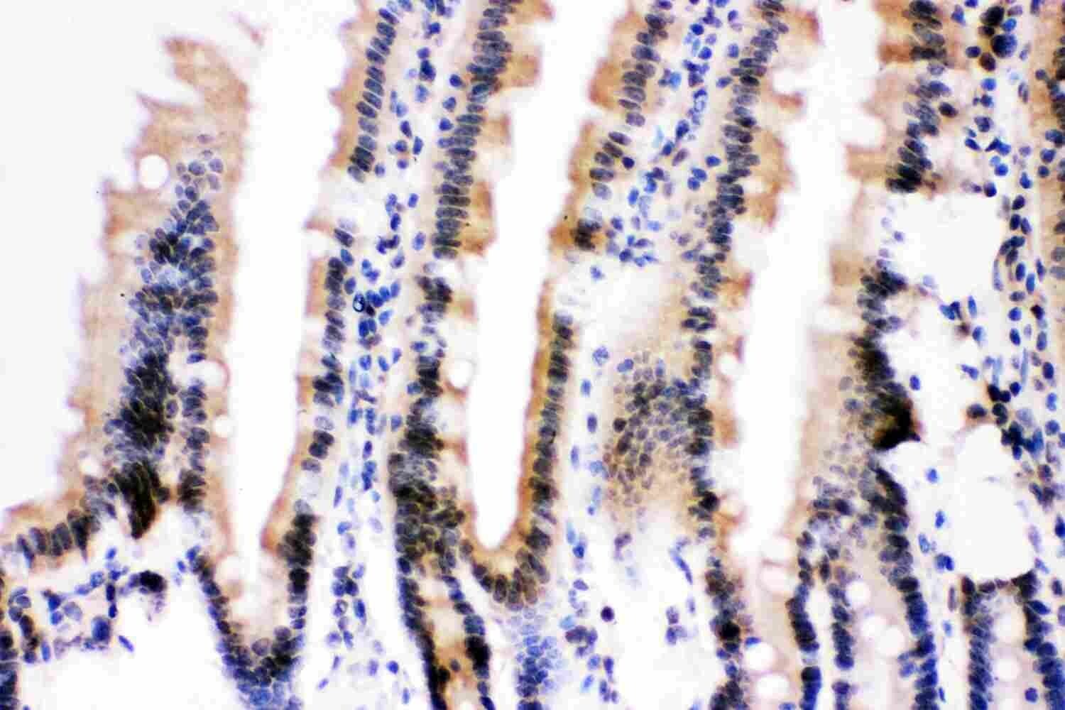
- Immunohistochemistry analysis of Ubiquitin B in paraffin-embedded rat intestine tissue. Antigen retrieval was performed on the tissue using citrate buffer (pH 6, 20 min) and blocked with 10% goat serum. Samples were incubated with Ubiquitin B polyclonal antibody (Product # PA5-95195) at a 1 µg/mL dilution, followed by biotinylated goat anti-rabbit IgG (30 min, 37°C), and developed with Strepavidin-Biotin-Complex and DAB.

- Submitted by
- Invitrogen Antibodies (provider)
- Main image

- Experimental details
- Immunohistochemistry analysis of Ubiquitin B in paraffin-embedded human mammary cancer tissue. Antigen retrieval was performed on the tissue using citrate buffer (pH 6, 20 min) and blocked with 10% goat serum. Samples were incubated with Ubiquitin B polyclonal antibody (Product # PA5-95195) at a 1 µg/mL dilution, followed by biotinylated goat anti-rabbit IgG (30 min, 37°C), and developed with Strepavidin-Biotin-Complex and DAB.

- Submitted by
- Invitrogen Antibodies (provider)
- Main image

- Experimental details
- Immunohistochemistry analysis of Ubiquitin B in paraffin-embedded mouse intestine tissue. Antigen retrieval was performed on the tissue using citrate buffer (pH 6, 20 min) and blocked with 10% goat serum. Samples were incubated with Ubiquitin B polyclonal antibody (Product # PA5-95195) at a 1 µg/mL dilution, followed by biotinylated goat anti-rabbit IgG (30 min, 37°C), and developed with Strepavidin-Biotin-Complex and DAB.
Supportive validation
- Submitted by
- Invitrogen Antibodies (provider)
- Main image

- Experimental details
- Immunohistochemical localization of ubiquitin B in Eulalia . ( A ) Alignment of peptides from MS/MS on fluorescent SDS-PAGE gel bands, RNAseq sequences with positive matches to ubiquitin, and the human ubiquitin B sequence against which the polyclonal antibody was used. The antibody was chosen based on similarity with Eulalia ubiquitin isoform 3. ( B ) Paraffin section across a parapodium of the worm fixed with Zenker and stained with HE (hematoxylin and eosin). Hemocytes are identified by the arrows. Inset: Paraffin section fixed with glutaraldehyde and stained with TC (tetrachrome stain) showing hepatocytes naturally colored by green pigments. ( C ) Compositive of a cryopreserved section of a parapodia marked for ubiquitin B. ( D ) Negative control of cryopreserved section of a parapodia. ( E ) Paraffin section fixed with Zenker and stained with HE; arrows are indicative of stem cells. ( F ) Composite of a paraffin section of the ventral lateral section marked for ubiquitin B. Arrows indicate the stem cells marked by the antibody. ( G ) Negative control of cryopreserved section of the ventral lateral area, close to the parapodia. ( H ) Paraffin section fixed with glutaraldehyde and stained with HE, showing the female gametes in the celomic cavity. ( I ) Composite of celomic cavity with positive signal for ubiquitin B (arrow). ( J ) Negative control of cryopreserved section of the celomic cavity.
