Antibody data
- Antibody Data
- Antigen structure
- References [4]
- Comments [0]
- Validations
- Western blot [1]
- Immunohistochemistry [11]
- Flow cytometry [1]
Submit
Validation data
Reference
Comment
Report error
- Product number
- GTX16669 - Provider product page

- Provider
- GeneTex
- Proper citation
- GeneTex Cat#GTX16669, RRID:AB_425123
- Product name
- CD3 antibody [SP7]
- Antibody type
- Monoclonal
- Reactivity
- Human, Mouse, Rat, Canine, Feline, Horse, Porcine, Rabbit, Sheep, Simian
- Host
- Rabbit
Submitted references Evaluation of Explant Responses to STING Ligands: Personalized Immunosurgical Therapy for Head and Neck Squamous Cell Carcinoma.
In vivo amelioration of endogenous antitumor autoantibodies via low-dose P4N through the LTA4H/activin A/BAFF pathway.
Pathogenic aquaporin-4 reactive T cells are sufficient to induce mouse model of neuromyelitis optica.
Robust shifts in S100a9 expression with aging: a novel mechanism for chronic inflammation.
Baird JR, Bell RB, Troesch V, Friedman D, Bambina S, Kramer G, Blair TC, Medler T, Wu Y, Sun Z, de Gruijl TD, van de Ven R, Leidner RS, Crittenden MR, Gough MJ
Cancer research 2018 Nov 1;78(21):6308-6319
Cancer research 2018 Nov 1;78(21):6308-6319
In vivo amelioration of endogenous antitumor autoantibodies via low-dose P4N through the LTA4H/activin A/BAFF pathway.
Lin YL, Tsai NM, Hsieh CH, Ho SY, Chang J, Wu HY, Hsu MH, Chang CC, Liao KW, Jackson TL, Mold DE, Huang RC
Proceedings of the National Academy of Sciences of the United States of America 2016 Nov 29;113(48):E7798-E7807
Proceedings of the National Academy of Sciences of the United States of America 2016 Nov 29;113(48):E7798-E7807
Pathogenic aquaporin-4 reactive T cells are sufficient to induce mouse model of neuromyelitis optica.
Jones MV, Huang H, Calabresi PA, Levy M
Acta neuropathologica communications 2015 May 21;3:28
Acta neuropathologica communications 2015 May 21;3:28
Robust shifts in S100a9 expression with aging: a novel mechanism for chronic inflammation.
Swindell WR, Johnston A, Xing X, Little A, Robichaud P, Voorhees JJ, Fisher G, Gudjonsson JE
Scientific reports 2013;3:1215
Scientific reports 2013;3:1215
No comments: Submit comment
Supportive validation
- Submitted by
- GeneTex (provider)
- Main image

- Experimental details
- Western Blot analysis of Jurkat cell lysate with CD3 antibody
Supportive validation
- Submitted by
- GeneTex (provider)
- Main image

- Experimental details
- Formalin fixed paraffin embedded human tonsil stained with CD3antibody (GTX16669)
- Submitted by
- GeneTex (provider)
- Main image
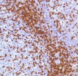
- Experimental details
- Human Tonsil stained with anti-CD3 antibody
- Submitted by
- GeneTex (provider)
- Main image
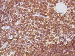
- Experimental details
- Human Thymus stained with anti-CD3 antibody
- Submitted by
- GeneTex (provider)
- Main image
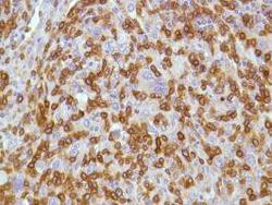
- Experimental details
- Human HK Lymphoma stained with anti-CD3 antibody
- Submitted by
- GeneTex (provider)
- Main image
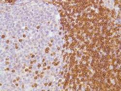
- Experimental details
- Human Tonsil stained with anti-CD3 antibody
- Submitted by
- GeneTex (provider)
- Main image
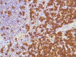
- Experimental details
- Human Reactive Lymph Node stained with anti-CD3 antibody
- Submitted by
- GeneTex (provider)
- Main image

- Experimental details
- Human Cervix stained with anti-CD3 antibody
- Submitted by
- GeneTex (provider)
- Main image
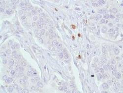
- Experimental details
- Human Breast Ductal Carcinoma stained with anti-CD3 antibody
- Submitted by
- GeneTex (provider)
- Main image
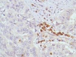
- Experimental details
- Human Lung Squamous Cell Carcinoma stained with anti-CD3 antibody
- Submitted by
- GeneTex (provider)
- Main image
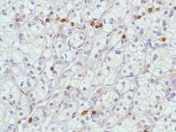
- Experimental details
- Human Renal Cell Carcinoma stained with anti-CD3 antibody
- Submitted by
- GeneTex (provider)
- Main image
- Experimental details
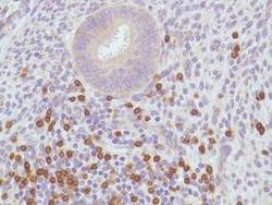
- Human Uterus stained with anti-CD3 antibody
Supportive validation
- Submitted by
- GeneTex (provider)
- Main image
- Experimental details
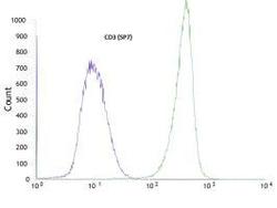
- Flow cytometric analysis of rabbit anti-CD3 (SP7) antibody in Jurkat (green) compare to negative control of rabbit IgG (blue)
 Explore
 Validate
 Learn
Learn Western blot