 Explore
 Validate
 Learn
Learn Western blot
Antibody data
- Antibody Data
- Antigen structure
- References [1]
- Comments [0]
- Validations
- Western blot [2]
- Immunocytochemistry [2]
- Immunohistochemistry [9]
Submit
Validation data
Reference
Comment
Report error
- Product number
- HPA035636 - Provider product page

- Provider
- Atlas Antibodies
- Proper citation
- Atlas Antibodies Cat#HPA035636, RRID:AB_10670837
- Product name
- Anti-AARS2
- Antibody type
- Polyclonal
- Description
- Polyclonal Antibody against Human AARS2, Gene description: alanyl-tRNA synthetase 2, mitochondrial, Alternative Gene Names: AARSL, bA444E17.1, KIAA1270, Validated applications: ICC, IHC, WB, Uniprot ID: Q5JTZ9, Storage: Store at +4°C for short term storage. Long time storage is recommended at -20°C.
- Reactivity
- Human
- Host
- Rabbit
- Conjugate
- Unconjugated
- Isotype
- IgG
- Vial size
- 100 µl
- Concentration
- 0.2 mg/ml
- Storage
- Store at +4°C for short term storage. Long time storage is recommended at -20°C.
Submitted references Editing activity for eliminating mischarged tRNAs is essential in mammalian mitochondria
Hilander T, Zhou X, Konovalova S, Zhang F, Euro L, Chilov D, Poutanen M, Chihade J, Wang E, Tyynismaa H
Nucleic Acids Research 2018;46(2):849-860
Nucleic Acids Research 2018;46(2):849-860
No comments: Submit comment
Supportive validation
- Submitted by
- Atlas Antibodies (provider)
- Main image
- Experimental details
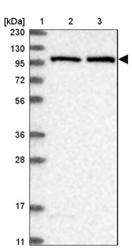
- Lane 1: Marker [kDa] 230, 130, 95, 72, 56, 36, 28, 17, 11Lane 2: Human cell line RT-4Lane 3: Human cell line U-251MG sp

- Submitted by
- Atlas Antibodies (provider)
- Main image

- Experimental details
- Western blot analysis in human cell line SCLC-21H.
- Sample type
- Human
- Protocol
- Protocol

Supportive validation
- Submitted by
- Atlas Antibodies (provider)
- Main image

- Experimental details
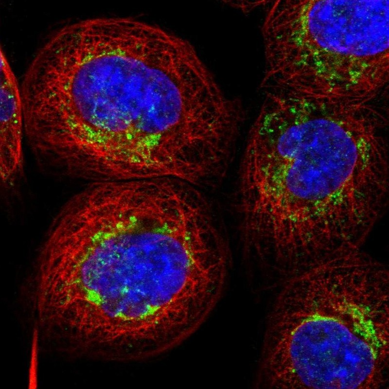
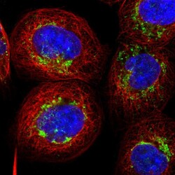
- Immunofluorescent staining of human cell line A-431 shows localization to mitochondria.
- Sample type
- HUMAN
- Submitted by
- Atlas Antibodies (provider)
- Main image
- Experimental details
- Immunofluorescent staining of human cell line A-431 shows localization to mitochondria.
- Sample type
- Human
- Protocol
- Protocol

Supportive validation
- Submitted by
- Atlas Antibodies (provider)
- Main image
- Experimental details
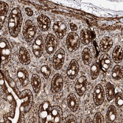
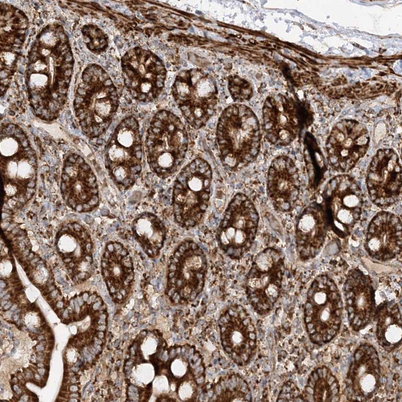
- Immunohistochemical staining of human duodenum shows strong cytoplasmic positivity in glandular cells.
- Sample type
- HUMAN
- Submitted by
- Atlas Antibodies (provider)
- Main image
- Experimental details
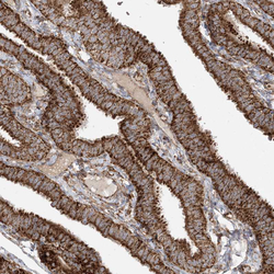
- Immunohistochemical staining of human Fallopian tube shows strong granular cytoplasmic positivity in glandular cells.
- Sample type
- HUMAN

- Submitted by
- Atlas Antibodies (provider)
- Main image

- Experimental details
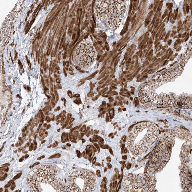
- Immunohistochemical staining of human prostate shows strong cytoplasmic positivity in smooth muscle cells.
- Sample type
- HUMAN
- Submitted by
- Atlas Antibodies (provider)
- Main image

- Experimental details
- Immunohistochemical staining of human duodenum shows strong granular cytoplasmic positivity in glandular cells.
- Sample type
- HUMAN
- Submitted by
- Atlas Antibodies (provider)
- Main image
- Experimental details
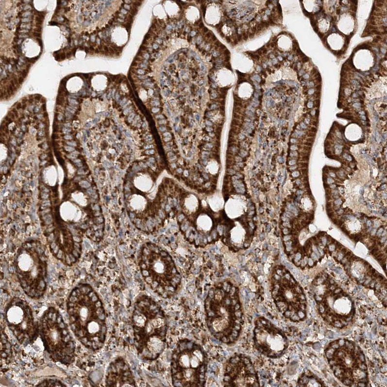
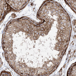
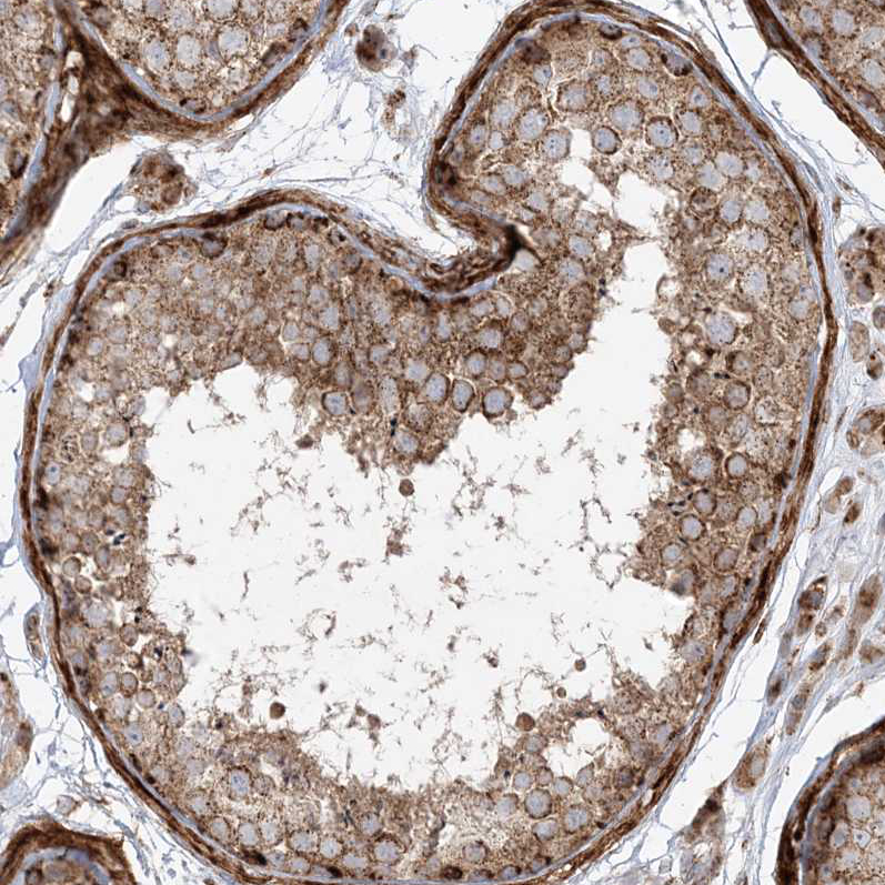
- Immunohistochemical staining of human testis shows moderate to strong granular cytoplasmic positivity in cells in seminiferous ducts.
- Sample type
- HUMAN
- Submitted by
- Atlas Antibodies (provider)
- Main image

- Experimental details
- Immunohistochemical staining of human duodenum shows strong granular cytoplasmic positivity in glandular cells.
- Sample type
- Human
- Protocol
- Protocol

- Submitted by
- Atlas Antibodies (provider)
- Main image

- Experimental details
- Immunohistochemical staining of human Fallopian tube shows strong granular cytoplasmic positivity in glandular cells.
- Sample type
- Human
- Protocol
- Protocol

- Submitted by
- Atlas Antibodies (provider)
- Main image

- Experimental details
- Immunohistochemical staining of human prostate shows strong cytoplasmic positivity in smooth muscle cells.
- Sample type
- Human
- Protocol
- Protocol

- Submitted by
- Atlas Antibodies (provider)
- Main image

- Experimental details
- Immunohistochemical staining of human testis shows moderate to strong granular cytoplasmic positivity in cells in seminiferous ducts.
- Sample type
- Human
- Protocol
- Protocol
