Explore
 Validate
 Learn
Learn Western blot
Antibody data
- Antibody Data
- Antigen structure
- References [0]
- Comments [0]
- Validations
- Western blot [2]
- Immunocytochemistry [1]
- Immunohistochemistry [1]
Submit
Validation data
Reference
Comment
Report error
- Product number
- TA328975 - Provider product page

- Provider
- OriGene
- Product name
- Rabbit polyclonal Anti-K2P18.1 (TRESK) (extracellular)
- Antibody type
- Polyclonal
- Description
- Rabbit polyclonal Anti-K2P18.1 (TRESK) (extracellular)
- Host
- Rabbit
- Conjugate
- Unconjugated
- Epitope
- Kcnk18
- Antibody clone number
- NULL
- Vial size
- 200 µl
- Concentration
- NULL
No comments: Submit comment
Supportive validation
- Submitted by
- OriGene (provider)
- Main image
- Experimental details
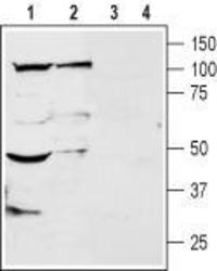
- Western blot analysis of rat brain (lanes 1 and 3) and dorsal root ganglion (lanes 2 and 4) membranes: 1, 2. Anti-K2P18.1 (TRESK) (extracellular) antibody , (1:200). 3, 4. Anti-K2P18.1 (TRESK) (extracellular), preincubated with the control peptide antigen.
- Validation comment
- WB
- Submitted by
- OriGene (provider)
- Main image

- Experimental details
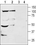
- Western blot analysis of mouse brain membranes: 1. Anti-K2P18.1 (TRESK) (extracellular) antibody , (1:200). 2. Anti-K2P18.1 (TRESK) (extracellular) antibody, preincubated with the control peptide antigen.
- Validation comment
- WB
Supportive validation
- Submitted by
- OriGene (provider)
- Main image
- Experimental details
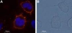
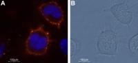
- Expression of K2P18.1 in ND7/23 cell line. Immunocytochemical staining of K2P18.1 channel in a mouse/rat neuroblastoma x dorsal root ganglion neuron hybrid cell line (ND7/23). A. Cells were stained with Anti- K2P18.1 (TRESK) (extracellular) antibody , (1:50) followed by goat-anti-rabbit-AlexaFluor-555 secondary antibody (red). Nuclei were visualized with the cell permeable dye Hoechst 33342 (blue staining). B. Show visible light images of the cells shown on (A).
- Validation comment
- IF
Supportive validation
- Submitted by
- OriGene (provider)
- Main image
- Experimental details
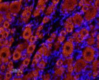
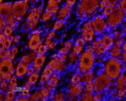
- Expression of K2P18.1 in rat dorsal root ganglia. Immunohistochemical staining of rat dorsal root ganglia (DRG) frozen sections using Anti- K2P18.1 (TRESK) (extracellular) antibody , (1:100) followed by Alexa 555-labeled secondary antibody (red staining). Both big/medium sized neurons and small neurons are stained. Note that glial cells and axonal fibers are not stained. Hoechst 33342 is used as the counterstain (blue staining).
- Validation comment
- IHC