Explore
 Validate
 Learn
Learn Western blot
 Immunoprecipitation
Antibody data
- Antibody Data
- Antigen structure
- References [0]
- Comments [0]
- Validations
- Western blot [1]
- Other assay [2]
Submit
Validation data
Reference
Comment
Report error
- Product number
- PA5-112063 - Provider product page

- Provider
- Invitrogen Antibodies
- Product name
- LILRA1 Polyclonal Antibody
- Antibody type
- Polyclonal
- Antigen
- Synthetic peptide
- Reactivity
- Human
- Host
- Rabbit
- Isotype
- IgG
- Vial size
- 100 µL
- Concentration
- 1 mg/mL
- Storage
- Store at 4°C short term. For long term storage, store at -20°C, avoiding freeze/thaw cycles.
No comments: Submit comment
Supportive validation
- Submitted by
- Invitrogen Antibodies (provider)
- Main image

- Experimental details
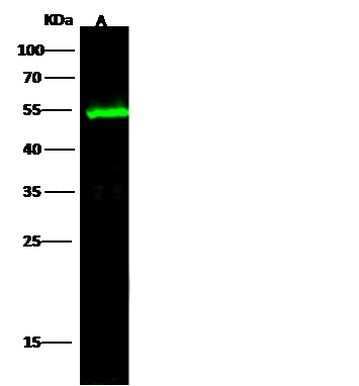
- Western Blot analysis of LILRA1 using LILRA1 Polyclonal Antibody (Product # PA5-112063) at a dilution of 1:500. Lane A: MCF7 Membrane Lysate , (Lysates/proteins at 30 µg per lane). The secondary antibody used was a Goat Anti-Rabbit IgG H&L (Dylight800) at 1:10000 dilution. Developed using the Odyssey technique and performed under reducing conditions. Predicted band size: 53 kDa. Observed band size: 53 kDa.
Supportive validation
- Submitted by
- Invitrogen Antibodies (provider)
- Main image
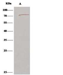
- Experimental details
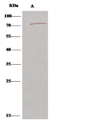
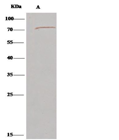
- Immunoprecipitation of LILRA1 was performed on (Lane A) 0.5 mg MCF-7 whole cell lysate using 2 µL of LILRA1 Polyclonal Antibody (Product # PA5-112063) at a dilution of 1:100, and 15 µL of 50 % Protein G agarose. A Clean Blot IP Detection Reagent (HRP) was used as a secondary antibody at a dilution of 1:1000. Developed using the DAB staining technique and performed under reducing conditions. Predicted band size: 53 kDa. Observed band size: 70 kDa.
- Submitted by
- Invitrogen Antibodies (provider)
- Main image
- Experimental details
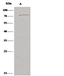
- Immunoprecipitation of LILRA1 was performed on (Lane A) 0.5 mg MCF-7 whole cell lysate using 2 µL of LILRA1 Polyclonal Antibody (Product # PA5-112063) at a dilution of 1:100, and 15 µL of 50 % Protein G agarose. A Clean Blot IP Detection Reagent (HRP) was used as a secondary antibody at a dilution of 1:1000. Developed using the DAB staining technique and performed under reducing conditions. Predicted band size: 53 kDa. Observed band size: 70 kDa.