Explore
 Validate
 Learn
Learn Western blot
Antibody data
- Antibody Data
- Antigen structure
- References [0]
- Comments [0]
- Validations
- Western blot [2]
- Immunohistochemistry [2]
- Flow cytometry [1]
Submit
Validation data
Reference
Comment
Report error
- Product number
- ACL-031-200UL - Provider product page

- Provider
- Invitrogen Antibodies
- Product name
- PACC1/TMEM206 (extracellular) Polyclonal Antibody
- Antibody type
- Polyclonal
- Antigen
- Other
- Description
- Reconstitution: 1 X 25 µL double distilled water (DDW), depending on the sample size. The antibody ships as a lyophilized powder at room temperature. Upon arrival, it should be stored at -20C. The reconstituted solution can be stored at 4C, protected from the light, for up to 1 week. For longer periods, small aliquots should be stored at -20C. Avoid multiple freezing and thawing. Centrifuge all antibody preparations before use (10000 X g 5 min).
- Reactivity
- Human, Mouse, Rat
- Host
- Rabbit
- Isotype
- IgG
- Vial size
- 200 µL
- Concentration
- 0.8 mg/mL
- Storage
- -20° C, Avoid Freeze/Thaw Cycles
No comments: Submit comment
Supportive validation
- Submitted by
- Invitrogen Antibodies (provider)
- Main image

- Experimental details
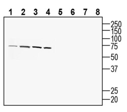
- Western blot analysis of rat brain lysate (lanes 1 and 4), mouse brain membranes (lanes 2 and 5) and rat kidney lysates (lanes 3 and 6): - 1-3. Anti-PACC1/TMEM206 (extracellular) Antibody (#ACL-031), (1:200). 4-6. Anti-PACC1/TMEM206 (extracellular) Antibody , preincubated with PACC1/TMEM206 (extracellular) Blocking Peptide (BLP-CL031).

- Submitted by
- Invitrogen Antibodies (provider)
- Main image
- Experimental details
- Western blot analysis of human U-87 MG glioblastoma cell line lysate (lanes 1 and 5), human HT-29 colon adenocarcinoma cell line lysate (lanes 2 and 6), human THP-1 monocytic leukemia cell line lysate (lanes 3 and 7) and mouse BV-2 microglia cell line lysate (lanes 4 and 8): - 1-4. Anti-PACC1/TMEM206 (extracellular) Antibody (#ACL-031), (1:200). 5-8. Anti-PACC1/TMEM206 (extracellular) Antibody , preincubated with PACC1/TMEM206 (extracellular) Blocking Peptide (BLP-CL031).

Supportive validation
- Submitted by
- Invitrogen Antibodies (provider)
- Main image
- Experimental details
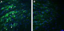
- Expression of PACC1 in mouse reticular thalamic nucleus. Immunohistochemical staining of perfusion-fixed frozen mouse brain sections with Anti-PACC1/TMEM206 (extracellular) Antibody (#ACL-031), (1:200), followed by goat Anti-rabbit-AlexaFluor-488. A. PACC1 immunoreactivity (green) appears in neurons (arrows). B. Pre-incubation of the Antibody with PACC1/TMEM206 (extracellular) Blocking Peptide (BLP-CL031), suppressed staining. Cell nuclei are stained with DAPI (blue).

- Submitted by
- Invitrogen Antibodies (provider)
- Main image
- Experimental details
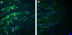
- Expression of PACC1 in mouse reticular thalamic nucleus. Immunohistochemical staining of perfusion-fixed frozen mouse brain sections with Anti-PACC1/TMEM206 (extracellular) Antibody (#ACL-031), (1:200), followed by goat Anti-rabbit-AlexaFluor-488. A. PACC1 immunoreactivity (green) appears in neurons (arrows). B. Pre-incubation of the Antibody with PACC1/TMEM206 (extracellular) Blocking Peptide (BLP-CL031), suppressed staining. Cell nuclei are stained with DAPI (blue).

Supportive validation
- Submitted by
- Invitrogen Antibodies (provider)
- Main image
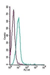
- Experimental details
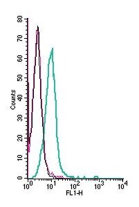
- Cell surface detection of PACC1 by indirect flow cytometry in live intact human THP-1 monocytic leukemia cell line: - (black line) cells. (red) Cells + goat- Anti-rabbit-FITC. (green) Cells + Anti-PACC1/TMEM206 (extracellular) Antibody (#ACL-031), (2.5μg) + goat- Anti-rabbit-FITC.