 Explore
 Validate
 Learn
Learn Western blot
Antibody data
- Antibody Data
- Antigen structure
- References [1]
- Comments [0]
- Validations
- Western blot [4]
- Immunocytochemistry [1]
- Immunohistochemistry [2]
- Other assay [1]
Submit
Validation data
Reference
Comment
Report error
- Product number
- PA5-27150 - Provider product page

- Provider
- Invitrogen Antibodies
- Product name
- GPX2 Polyclonal Antibody
- Antibody type
- Polyclonal
- Antigen
- Synthetic peptide
- Description
- Recommended positive controls: HepG2.
- Concentration
- 1.39 mg/mL
Submitted references Clinicopathological and prognostic significance of GPX2 protein expression in esophageal squamous cell carcinoma.
Lei Z, Tian D, Zhang C, Zhao S, Su M
BMC cancer 2016 Jul 7;16:410
BMC cancer 2016 Jul 7;16:410
No comments: Submit comment
Supportive validation
- Submitted by
- Invitrogen Antibodies (provider)
- Main image

- Experimental details
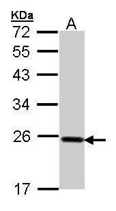
- Western blot analysis of GPX2 using 30 µg of HepG2 lysate. Samples were loaded onto a 12% SDS-PAGE gel and probed with a GPX2 polyclonal antibody (Product # PA5-27150) at a dilution of 1:1000.
- Submitted by
- Invitrogen Antibodies (provider)
- Main image
- Experimental details
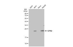
- Western Blot using GPX2 Polyclonal Antibody (Product # PA5-27150). Various whole cell extracts (30 µg) were separated by 12% SDS-PAGE, and the membrane was blotted with GPX2 Polyclonal Antibody (Product # PA5-27150) diluted at 1:1,000. The HRP-conjugated anti-rabbit IgG antibody was used to detect the primary antibody.

- Submitted by
- Invitrogen Antibodies (provider)
- Main image

- Experimental details
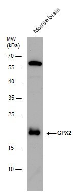
- GPX2 antibody detects GPX2 protein by Western blot analysis. Mouse tissue extracts (50 µg) was separated by 12 % SDS-PAGE, and blotted with GPX2 antibody GPX2 Polyclonal Antibody (Product # PA5-27150) diluted by 1:500.
- Submitted by
- Invitrogen Antibodies (provider)
- Main image

- Experimental details
- GPX2 Polyclonal Antibody detects GPX2 protein by Western blot analysis. Whole cell extracts (30 µg) was separated by 12 % SDS-PAGE, and blotted with GPX2 Polyclonal Antibody (Product # PA5-27150) diluted by 1:1,000.

Supportive validation
- Submitted by
- Invitrogen Antibodies (provider)
- Main image

- Experimental details
- GPX2 Polyclonal Antibody detects GPX2 protein at granule by immunofluorescent analysis. Sample: HepG2 cells were fixed in 4% paraformaldehyde at RT for 15 min. Green: GPX2 stained by GPX2 Polyclonal Antibody (Product # PA5-27150) diluted at 1:500. Red: alpha Tubulin, a cytoskeleton marker, stained by alpha Tubulin Polyclonal Antibody [GT114] (Product # MA5-31466) diluted at 1:1,000. Blue: Fluoroshield with DAPI .

Supportive validation
- Submitted by
- Invitrogen Antibodies (provider)
- Main image

- Experimental details
- Immunohistochemical analysis of paraffin-embedded human gastric cancer, using GPX2 (Product # PA5-27150) antibody at 1:500 dilution. Antigen Retrieval: EDTA based buffer, pH 8.0, 15 min.

- Submitted by
- Invitrogen Antibodies (provider)
- Main image
- Experimental details
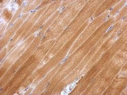
- Immunohistochemical analysis of paraffin-embedded mouse muscle, using GPX2 (Product # PA5-27150) antibody at 1:500 dilution. Antigen Retrieval: EDTA based buffer, pH 8.0, 15 min.

Supportive validation
- Submitted by
- Invitrogen Antibodies (provider)
- Main image
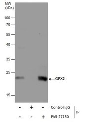
- Experimental details
- Immunoprecipitation of GPX2 was performed in HepG2 whole cell extracts using 5 µg of GPX2 Polyclonal Antibody (Product # PA5-27150). Samples were transferred to a membrane and probed with GPX2 Polyclonal Antibody as a primary antibody and an HRP-conjugated anti-Rabbit IgG was used as a secondary antibody.
